Clinical Assessment & Protocol
Typical Presentation (HPI)
EN: A 65-year-old patient reports worsening knee pain with activity and stiffness after sitting. AR: مريض يبلغ من العمر 65 عاماً يبلغ عن تفاقم ألم الركبة مع النشاط وتيبس بعد الجلوس.
General Examination
EN: Unremarkable or not routinely indicated. AR: طبيعي أو غير مطلوب روتينياً.
Treatment Protocol
EN: Strength training, weight management, and hyaluronic acid injections. AR: تدريبات القوة، إدارة الوزن، وحقن حمض الهيالورونيك.
Patient Education
EN: Low-impact aerobic exercises such as swimming or cycling. AR: تمارين هوائية منخفضة التأثير مثل السباحة أو ركوب الدراجات.
Systemic & Specialized Examinations
EN: S1, S2 present. No murmurs. AR: صوتا القلب الأول والثاني طبيعيان. لا توجد نفخات.
EN: Lungs clear to auscultation. AR: الرئتان صافيتان عند التسمع.
EN: Abdomen soft, non-tender. AR: البطن لين ولا يوجد ألم.
EN: Alert, oriented x3. No focal deficits. AR: المريض واعي ومدرك. لا يوجد عجز عصبي بؤري.
EN: Unremarkable or not routinely indicated. AR: طبيعي أو غير مطلوب روتينياً.
EN: Unremarkable or not routinely indicated. AR: طبيعي أو غير مطلوب روتينياً.
EN: Unremarkable or not routinely indicated. AR: طبيعي أو غير مطلوب روتينياً.
EN: Unremarkable or not routinely indicated. AR: طبيعي أو غير مطلوب روتينياً.
EN: Unremarkable or not routinely indicated. AR: طبيعي أو غير مطلوب روتينياً.
Orthopedic & Trauma Assessments
EN: Unremarkable or not routinely indicated. AR: طبيعي أو غير مطلوب روتينياً.
EN: Crepitus on range of motion, medial joint line tenderness, and bony enlargement. AR: فرقعة عند تحريك المفصل، إيلام عند خط المفصل الإنسي، وتضخم عظمي.
Comprehensive Clinical Guide: Osteoarthritis of the Knee (Gonarthrosis)
1. Introduction and Overview
Osteoarthritis (OA) of the knee, clinically referred to as gonarthrosis, is a chronic, degenerative joint disease characterized by the progressive deterioration of articular cartilage, subchondral bone remodeling, osteophyte formation, and synovial inflammation. It is the most common form of arthritis globally and a leading cause of disability, particularly in aging populations.
Unlike inflammatory arthropathies (such as Rheumatoid Arthritis), gonarthrosis is primarily a "wear-and-tear" condition, though current research confirms it is a metabolically active disease process involving the entire joint organ. As the articular cartilage thins, the mechanical environment of the knee shifts, leading to joint space narrowing (JSN), pain, stiffness, and eventual loss of function.
2. Etiology and Pathophysiology
The Mechanism of Degeneration
The pathophysiology of gonarthrosis is multifactorial. It is no longer viewed as a simple mechanical erosion but rather an imbalance between cartilage synthesis and degradation.
- Chondrocyte Dysfunction: Chondrocytes (the cells responsible for maintaining the extracellular matrix) shift from an anabolic state to a catabolic state. They release matrix metalloproteinases (MMPs) and aggrecanases that degrade Type II collagen and proteoglycans.
- Subchondral Bone Remodeling: As cartilage thins, the underlying bone experiences increased mechanical stress. This triggers bone resorption and sclerosis, leading to the formation of subchondral cysts and osteophytes at the joint margins.
- Synovial Inflammation: Low-grade chronic inflammation (synovitis) is present in most OA cases, contributing to the production of pro-inflammatory cytokines such as IL-1β and TNF-α, which further accelerate cartilage breakdown.
Key Risk Factors
| Factor Type | Specific Elements |
|---|---|
| Non-Modifiable | Age (>50), Genetic predisposition, Female gender, Anatomy (valgus/varus deformity). |
| Modifiable | Obesity (increased load), History of joint injury (ACL/meniscus tears), Occupation (repetitive kneeling/lifting). |
3. Clinical Staging and Grading
The gold standard for staging gonarthrosis is the Kellgren-Lawrence (K-L) Grading System, based on radiographic findings.
| Grade | Description | Clinical Presentation |
|---|---|---|
| 0 | Normal | No radiographic features of OA. |
| 1 | Doubtful | Possible osteophytic lipping; doubtful joint space narrowing. |
| 2 | Mild | Definite osteophytes; possible joint space narrowing. |
| 3 | Moderate | Multiple osteophytes, definite JSN, some sclerosis. |
| 4 | Severe | Large osteophytes, marked JSN, severe sclerosis, bone deformity. |
4. Clinical Presentation and Diagnosis
Standard Presentation
Patients typically present with:
* Activity-related pain: Pain that worsens with movement and improves with rest.
* Morning stiffness: Usually lasting less than 30 minutes (distinguishing it from inflammatory arthritis).
* Crepitus: Audible or palpable grinding during range-of-motion (ROM) testing.
* Joint instability: A sensation of the knee "giving way."
* Reduced Range of Motion: Difficulty with deep flexion or full extension.
Diagnostic Testing
- Weight-Bearing Radiographs: AP, lateral, and sunrise views are essential to visualize joint space narrowing and osteophyte formation.
- MRI (Magnetic Resonance Imaging): Not required for routine diagnosis but highly effective for visualizing early cartilage defects, meniscal tears, and bone marrow lesions (BMLs).
- Laboratory Studies: Used primarily to rule out other conditions (e.g., Rheumatoid Factor, ESR, CRP, and synovial fluid analysis to rule out septic arthritis or gout).
5. Differential Diagnosis
It is critical to distinguish gonarthrosis from other knee pathologies:
* Rheumatoid Arthritis: Usually bilateral, symmetric, and characterized by systemic symptoms and morning stiffness >1 hour.
* Meniscal Injury: Often presents with locking/catching; usually acute onset compared to the insidious onset of OA.
* Patellofemoral Pain Syndrome: Common in younger patients; pain is typically anterior and related to prolonged sitting.
* Septic Arthritis: Acute, hot, swollen joint with fever; requires immediate aspiration.
6. Management and Therapeutic Approaches
Conservative Management (First-Line)
- Weight Loss: Every pound of weight loss reduces four pounds of pressure on the knee joint.
- Physical Therapy: Strengthening the quadriceps and hamstrings is the most effective way to stabilize the knee and reduce pain.
- Bracing/Orthotics: Unloader braces for unicompartmental OA; shoe inserts for malalignment.
Pharmacological Interventions
- NSAIDs: Topical (Diclofenac) is preferred to minimize systemic side effects; oral NSAIDs for acute flares.
- Intra-articular Injections: Corticosteroids for acute inflammation; Hyaluronic acid (viscosupplementation) for lubrication; PRP (Platelet-Rich Plasma) as an emerging biological therapy.
Surgical Interventions
- Arthroscopic Debridement: Generally discouraged for OA; indicated only for mechanical symptoms (locking).
- Osteotomy: Realignment surgery for younger, active patients with unicompartmental disease.
- Total Knee Arthroplasty (TKA): The definitive treatment for end-stage (K-L Grade 4) OA.
7. Risks, Side Effects, and Contraindications
- NSAID Risks: Chronic use carries risks of gastrointestinal bleeding, hypertension, and renal impairment.
- Corticosteroid Risks: Frequent intra-articular injections can lead to cartilage atrophy and increased infection risk.
- Surgical Risks: TKA carries risks of deep vein thrombosis (DVT), pulmonary embolism, periprosthetic infection, and chronic post-surgical pain.
8. Long-Term Prognosis
Gonarthrosis is a progressive disease. While no cure exists to "reverse" the cartilage loss, the prognosis is generally favorable with proactive management. Most patients maintain functional independence through a combination of lifestyle modifications and, when necessary, surgical intervention. Early diagnosis and weight management are the two most significant factors in slowing the progression of the disease.
9. Massive FAQ Section
1. Is knee osteoarthritis curable?
No, it is a degenerative condition. However, symptoms can be managed effectively, and functional mobility can be maintained for life.
2. Does running cause osteoarthritis?
Moderate running in healthy individuals is generally not associated with OA. However, elite-level impact loading or running on pre-existing joint injuries can accelerate degeneration.
3. When is the right time for knee replacement?
Surgery is typically considered when conservative measures (PT, weight loss, medication) fail to provide sufficient pain relief to perform daily activities.
4. What is the difference between "bone-on-bone" and normal OA?
"Bone-on-bone" refers to the complete loss of articular cartilage, where the femur and tibia make direct contact. This is typically K-L Grade 4.
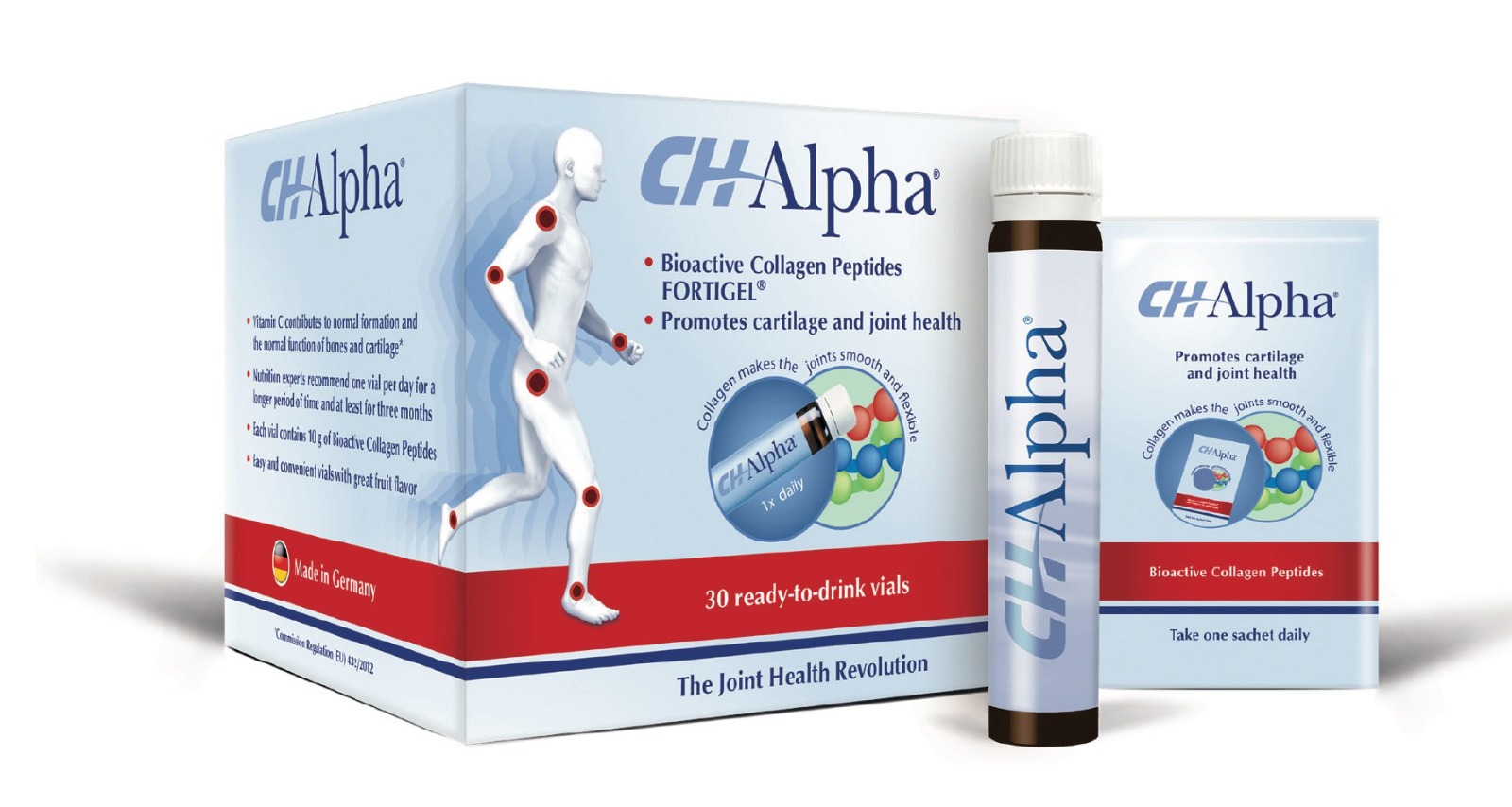
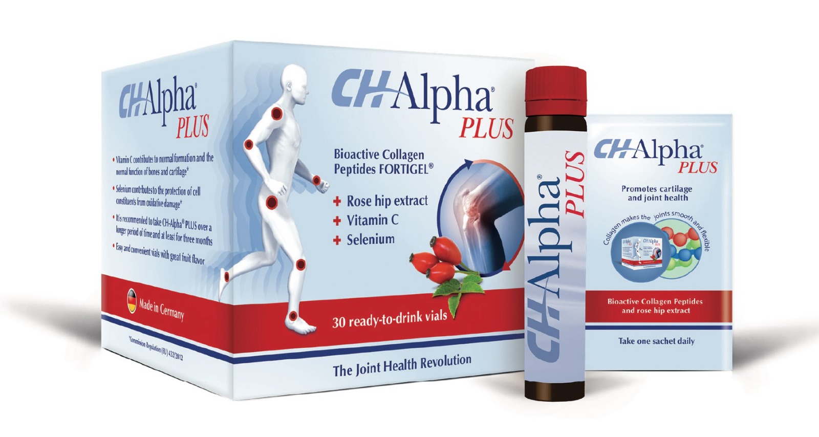
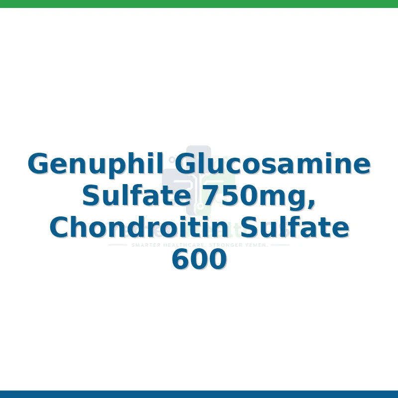
5. Can supplements like Glucosamine and Chondroitin help?
Clinical evidence is mixed. While some patients report relief, large-scale studies have not consistently proven these supplements to be superior to a placebo.
6. How long does a knee replacement last?
Modern implants are designed to last 15–25 years, depending on the patient's activity level and weight.
7. Is knee pain always osteoarthritis?
No. Pain can stem from bursitis, tendinitis, meniscal tears, or systemic arthritis. A clinical evaluation is necessary.
8. Why does my knee hurt more when it rains?
Changes in barometric pressure can cause expansion or contraction of the joint tissues, which can aggravate the sensitive nerve endings in a damaged joint.
9. Are knee braces helpful?
Yes, especially for those with varus or valgus deformity. "Unloader" braces shift the weight away from the damaged compartment of the knee.
10. How much weight do I need to lose to help my knees?
Even a 5% to 10% reduction in total body weight has been shown to significantly reduce pain and improve functional scores in patients with gonarthrosis.
10. Conclusion for Clinicians
Management of gonarthrosis requires a patient-centered approach. Clinicians should prioritize non-pharmacological interventions, emphasizing the "Big Three": weight management, physical therapy, and patient education. Surgical consultation should be reserved for those who have exhausted conservative avenues and whose quality of life is severely impacted by pain. By framing OA as a manageable chronic condition rather than an inevitable decline, providers can significantly improve patient outcomes and long-term joint health.