Clinical Assessment & Protocol
Typical Presentation (HPI)
EN: A 20-year-old volleyball player reports chronic anterior knee pain localized to the inferior pole of the patella. AR: لاعب كرة طائرة يبلغ من العمر 20 عاماً يشكو من ألم مزمن في مقدمة الركبة متركز في القطب السفلي للرضفة.
General Examination
EN: Point tenderness at the inferior patellar pole; pain on resisted extension. AR: إيلام موضعي عند القطب الرضفي السفلي؛ ألم عند مقاومة بسط الركبة.
Treatment Protocol
EN: Eccentric loading exercises, activity modification, and shockwave therapy. AR: تمارين التحميل اللامركزي، تعديل النشاط، والعلاج بالموجات التصادمية.
Patient Education
EN: Emphasize patience with long-term rehabilitation. AR: التأكيد على الصبر في عملية إعادة التأهيل طويلة الأمد.
Systemic & Specialized Examinations
EN: S1, S2 present. No murmurs. AR: صوتا القلب الأول والثاني طبيعيان. لا توجد نفخات.
EN: Lungs clear to auscultation. AR: الرئتان صافيتان عند التسمع.
EN: Abdomen soft, non-tender. AR: البطن لين ولا يوجد ألم.
EN: Alert, oriented x3. No focal deficits. AR: المريض واعي ومدرك. لا يوجد عجز عصبي بؤري.
EN: Unremarkable or not routinely indicated. AR: طبيعي أو غير مطلوب روتينياً.
EN: Unremarkable or not routinely indicated. AR: طبيعي أو غير مطلوب روتينياً.
EN: Unremarkable or not routinely indicated. AR: طبيعي أو غير مطلوب روتينياً.
EN: Unremarkable or not routinely indicated. AR: طبيعي أو غير مطلوب روتينياً.
EN: Unremarkable or not routinely indicated. AR: طبيعي أو غير مطلوب روتينياً.
Orthopedic & Trauma Assessments
EN: Unremarkable or not routinely indicated. AR: طبيعي أو غير مطلوب روتينياً.
EN: Unremarkable or not routinely indicated. AR: طبيعي أو غير مطلوب روتينياً.
Comprehensive Clinical Guide: Sports-Related Patellar Tendinopathy
Patellar tendinopathy, colloquially known as "Jumper’s Knee," represents one of the most prevalent and challenging overuse musculoskeletal pathologies encountered in sports medicine. Characterized by localized pain at the inferior pole of the patella, this condition frequently sidelines athletes involved in sports requiring repetitive, explosive loading of the extensor mechanism.
1. Clinical Definition and Overview
Patellar tendinopathy is a clinical syndrome involving structural degradation of the patellar tendon. Unlike inflammatory "tendinitis," which implies an acute inflammatory response, modern clinical consensus classifies this as a "tendinopathy"—a failure of the tendon’s healing response to repetitive mechanical overload, characterized by collagen disorganization, neovascularization, and cellular changes rather than classic inflammation.
It is most commonly identified in sports involving frequent jumping, landing, and rapid deceleration (e.g., volleyball, basketball, sprinting, and soccer).
2. Etiology and Pathophysiology
The Mechanism of Overload
The patellar tendon acts as the primary force transmitter between the quadriceps muscle group and the tibia. During high-intensity activity, the tendon is subjected to tensile, compressive, and shear forces.
- Tensile Forces: High-velocity eccentric loading during landing or deceleration.
- Compressive Forces: Impingement of the deep surface of the tendon against the inferior pole of the patella during knee flexion.
The Continuum Model of Tendinopathy
The prevailing model, proposed by Cook and Purdam, categorizes the pathology into three stages:
| Stage | Pathological Features | Clinical Implications |
|---|---|---|
| Reactive Tendinopathy | Non-inflammatory proliferative response to acute overload. | Potential for reversibility; tendon thickening. |
| Tendon Dysrepair | Increased matrix breakdown; neovascularization. | Structural changes; collagen disorganization. |
| Degenerative Tendinopathy | Cell death, large-scale matrix disruption, collagen separation. | Poor reversibility; high risk of rupture. |
Cellular and Structural Changes
- Hypercellularity: Increase in tenocyte count and fibroblast activity.
- Ground Substance Accumulation: Increased glycosaminoglycans (GAGs) leading to tendon swelling.
- Neovascularization: Ingrowth of blood vessels accompanied by sensory nerve fibers (the primary source of pain).
- Collagen Disarray: Shift from Type I (organized) to Type III (disorganized/weak) collagen.
3. Clinical Presentation and Staging
Standard Patient Presentation
Patients typically present with "warm-up" pain—discomfort that improves with initial activity but worsens significantly post-exercise or the following morning.
- Location: Pinpoint tenderness at the inferior pole of the patella (proximal tendon).
- Aggravating Factors: Squatting, jumping, stair climbing, and prolonged sitting (the "theater sign").
Blazina Staging System
The Blazina classification remains the gold standard for grading the severity of patellar tendinopathy:
| Stage | Symptom Description |
|---|---|
| I | Pain only after sports activity; no functional impairment. |
| II | Pain at the beginning of activity, disappears with warm-up, recurs after activity. |
| III | Pain during and after activity; patient unable to perform sports at a high level. |
| IV | Complete tendon rupture (surgical emergency). |
4. Diagnostic Evaluation
Physical Examination
The physical exam is the primary diagnostic tool.
* Palpation: Tenderness at the inferior patellar pole.
* Functional Testing: The Single-Leg Decline Squat (SLDS) is highly sensitive. The patient performs a squat on a 25-degree decline board; pain is typically provoked at 30–60 degrees of knee flexion.
* Quadriceps Flexibility: Assess for tightness, which increases tensile load on the tendon.
Imaging Modalities
- Ultrasound (US): Highly effective for identifying hypoechoic regions (degeneration), tendon thickening, and neovascularization (via Power Doppler).
- Magnetic Resonance Imaging (MRI): Gold standard for excluding intra-articular pathology (meniscal tears, chondromalacia). Shows high signal intensity on T2-weighted images.
- X-Ray: Generally used to rule out bone pathology, such as Osgood-Schlatter disease (in adolescents) or patellar spurring.
Differential Diagnosis
It is critical to distinguish patellar tendinopathy from other knee pathologies:
* Patellofemoral Pain Syndrome (PFPS): Diffuse peripatellar pain rather than localized tendon pain.
* Sinding-Larsen-Johansson Syndrome: Juvenile osteochondrosis of the inferior pole of the patella.
* Fat Pad Impingement (Hoffa’s Syndrome): Pain deep to the tendon, often associated with knee hyperextension.
* Patellar Bursitis: Swelling and tenderness anterior to the tendon, not within it.
5. Risks, Contraindications, and Management Considerations
Risks of Mismanagement
- Chronic Pain: Failure to address load management leads to irreversible degenerative changes.
- Tendon Rupture: Chronic degeneration significantly weakens the tendon, increasing the risk of full-thickness rupture during explosive movement.
- Muscle Atrophy: Prolonged rest without guided rehabilitation leads to quadriceps inhibition and atrophy.
Contraindications
- Corticosteroid Injections: Generally contraindicated in the patellar tendon due to the high risk of tendon weakening, collagen atrophy, and potential for rupture.
- Complete Immobilization: Leads to further collagen degradation and disuse atrophy.
6. Long-Term Prognosis and Rehabilitation
The prognosis is generally favorable for patients who adhere to a structured, progressive loading program. The core of treatment is Heavy Slow Resistance (HSR) training.
- Isometric Loading: Used for immediate pain relief (e.g., 5 sets of 45-second holds on a leg extension machine).
- Isotonic Loading: Concentric/eccentric loading to restore structural integrity.
- Energy Storage Loading: Reintroducing jumping and explosive movements in the final phase of rehab.
7. Frequently Asked Questions (FAQ)
1. Is "tendinitis" the same as "tendinopathy"?
No. Tendinitis implies acute inflammation. Tendinopathy is a degenerative condition involving cellular changes and collagen breakdown. Anti-inflammatories are often ineffective for the latter.
2. Can I continue to play sports while in pain?
"Pain-free" activity is rarely possible. Clinical guidelines suggest limiting activity to a level where pain is manageable (e.g., 3/10 on a visual analog scale) and does not persist the following morning.
3. Why do I feel pain when sitting for long periods?
This is known as the "theater sign." Prolonged knee flexion puts the patellar tendon under constant compressive load against the patella, triggering pain in sensitized tendons.
4. Are braces or straps effective?
Patellar tendon straps can provide symptomatic relief by changing the angle of force distribution, but they do not treat the underlying pathology. They should be used as an adjunct, not a cure.
5. How long does recovery take?
Recovery is rarely linear. A structured rehabilitation program typically requires 3 to 6 months to achieve significant functional improvement.
6. Should I get an MRI immediately?
Not necessarily. In the presence of classic clinical signs, MRI is often redundant. Imaging is reserved for cases that do not respond to 6–8 weeks of conservative therapy.
7. Can I use ice for the pain?
Ice may provide temporary analgesic relief, but it does not address the underlying degenerative changes. Use it sparingly for symptom management.
8. What is the role of PRP injections?
Platelet-Rich Plasma (PRP) is a biological therapy aimed at stimulating healing. Results are mixed, and it should only be considered after a failed 3–6 month course of conservative, load-based rehabilitation.
9. Is surgery ever required?
Surgery (e.g., debridement of the degenerative tissue) is a last resort, usually reserved for patients who have failed at least 6–12 months of high-quality, supervised physical therapy.
10. What is the best exercise for recovery?
Heavy Slow Resistance (HSR) training, particularly on a decline board, is currently the most evidence-supported method for stimulating tendon remodeling and increasing load-bearing capacity.
Conclusion
Sports-related patellar tendinopathy is a complex condition that demands a paradigm shift from passive "rest and ice" approaches to active, load-based management. By understanding the mechanical etiology and the stages of tendinopathy, clinicians can guide athletes through a safe return-to-sport protocol that emphasizes long-term tendon health over short-term symptomatic relief. Success in managing this condition requires patience, compliance with progressive loading, and a deep understanding of the patient's specific biomechanical vulnerabilities.
Related Clinical Integration
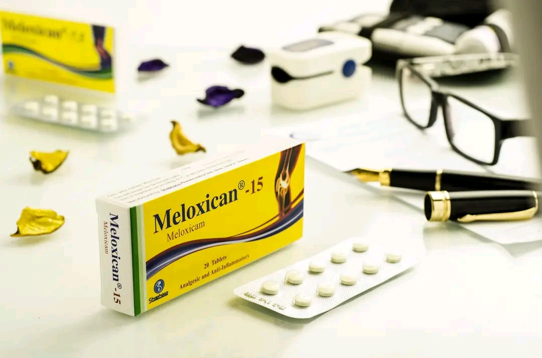
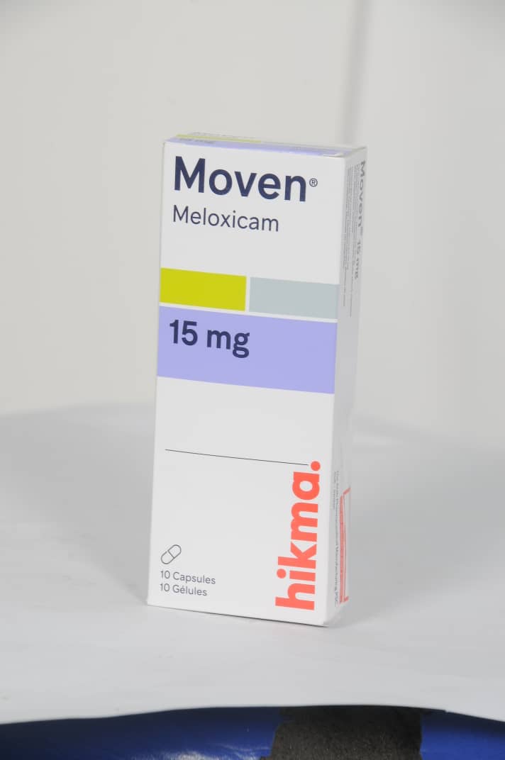
In the management of sports-related patellar tendinopathy, a multidisciplinary clinical approach is essential to address both acute inflammation and long-term functional recovery. Pharmacological intervention often involves the strategic use of non-steroidal anti-inflammatory drugs (NSAIDs) to manage pain and localized inflammation, including options such as Celcox / سيلكوكس 100mg, Celebrex / سيليبريكس 200mg, Diclofenac Sodium / ديكلوفيناك الصوديوم 75 mg, Ibuprofen / إيبوبروفين Not specified, Melflam 15mg 15mg, Meloxicam / ميلوكسيكام 25mg, Meloxican 15 mg, Mobic / موبيك 7.5mg, Mobic (Meloxicam) / موبيك (ميلوكسيكام) 7.5mg, Mobitil (Meloxicam) / موبيتيل (ميلوكسيكام) 15mg, Movalis / موفاليس 7.5mg, and [Moven15mg 30 capsules 15mg](https://yemenhealthos.com/ar/clinic/medications/moven15mg-30-