Clinical Assessment & Protocol
Typical Presentation (HPI)
EN: Itchy, scaly patches on the scalp with hair breakage. AR: بقع متقشرة ومثيرة للحكة في فروة الرأس مع تكسر الشعر.
General Examination
EN: Unremarkable or not routinely indicated. AR: طبيعي أو غير مطلوب روتينياً.
Treatment Protocol
EN: AR:
Patient Education
EN: AR:
Systemic & Specialized Examinations
EN: S1, S2 present. No murmurs. AR: صوتا القلب الأول والثاني طبيعيان. لا توجد نفخات.
EN: Lungs clear to auscultation. AR: الرئتان صافيتان عند التسمع.
EN: Abdomen soft, non-tender. AR: البطن لين ولا يوجد ألم.
EN: Alert, oriented x3. No focal deficits. AR: المريض واعي ومدرك. لا يوجد عجز عصبي بؤري.
EN: Scaling, alopecia, and 'black dot' hair remnants. AR: تقشر، ثعلبة، وبقايا شعر 'النقطة السوداء'.
EN: Unremarkable or not routinely indicated. AR: طبيعي أو غير مطلوب روتينياً.
EN: Unremarkable or not routinely indicated. AR: طبيعي أو غير مطلوب روتينياً.
EN: Unremarkable or not routinely indicated. AR: طبيعي أو غير مطلوب روتينياً.
EN: Unremarkable or not routinely indicated. AR: طبيعي أو غير مطلوب روتينياً.
Orthopedic & Trauma Assessments
EN: Unremarkable or not routinely indicated. AR: طبيعي أو غير مطلوب روتينياً.
EN: Unremarkable or not routinely indicated. AR: طبيعي أو غير مطلوب روتينياً.
1. Comprehensive Introduction & Overview
Tinea capitis, colloquially known as ringworm of the scalp, is a superficial dermatophytic fungal infection of the scalp hair follicles and the surrounding skin. While commonly diagnosed in pediatric populations, it represents a significant clinical challenge due to its potential for inflammatory complications, permanent alopecia, and social stigma.
As a primary dermatological condition, tinea capitis is classified by the site of infection and the nature of the fungal invasion. Unlike other forms of tinea (such as tinea corporis), tinea capitis involves the hair shaft, necessitating systemic antifungal therapy, as topical agents often fail to penetrate the hair follicle sufficiently to eradicate the fungal elements.
Epidemiological Context
The prevalence of tinea capitis has shifted significantly over the last century. Historically dominated by Microsporum audouinii, the current global landscape is heavily influenced by Trichophyton tonsurans in North America and Microsporum canis in Europe and parts of Asia. It predominantly affects prepubertal children, likely due to changes in sebum composition post-puberty that may possess antifungal properties.
2. Technical Specifications: Etiology and Pathophysiology
Etiological Agents
The pathogens responsible for tinea capitis are primarily anthropophilic, zoophilic, or geophilic fungi.
| Classification | Primary Pathogens | Source |
|---|---|---|
| Anthropophilic | Trichophyton tonsurans, M. audouinii | Human-to-human transmission |
| Zoophilic | Microsporum canis | Cats, dogs, farm animals |
| Geophilic | Microsporum gypseum | Soil contact |
Pathophysiological Mechanisms
The infection process follows a highly specific sequence:
1. Adherence: Fungal arthroconidia adhere to the stratum corneum of the scalp or the hair shaft.
2. Invasion: The fungus produces keratinases, allowing it to penetrate the hair cuticle.
3. Infection Pattern:
* Ectothrix: Fungal spores form a sheath around the outside of the hair shaft. The cuticle is destroyed.
* Endothrix: Fungal elements develop within the hair shaft, filling it with spores. The cuticle remains intact.
* Favus: The formation of scutula (crusts of hyphae and debris) leads to severe scarring.
3. Clinical Indications & Presentation
Tinea capitis presents with significant clinical heterogeneity, ranging from mild, non-inflammatory scaling to severe, pustular inflammatory responses.
Standard Clinical Presentations
- Non-inflammatory (Black Dot): Characterized by fine, grayish-white scaling of the scalp, often associated with broken-off hair shafts that appear as black dots at the follicular orifice.
- Inflammatory (Kerion): A severe, boggy, erythematous, and fluctuant plaque. This represents a hypersensitivity reaction to the fungal pathogen. It is often misdiagnosed as a bacterial abscess.
- Pustular/Seborrheic-like: Mimics seborrheic dermatitis with diffuse scaling and mild erythema.
Clinical Staging/Grading
While there is no universally standardized "stage" for tinea capitis, clinicians often categorize severity based on the inflammatory response:
* Grade I (Mild): Minimal scaling, no hair loss, no lymphadenopathy.
* Grade II (Moderate): Patchy alopecia, significant scaling, localized cervical lymphadenopathy.
* Grade III (Severe/Kerion): Boggy, exudative mass, diffuse lymphadenopathy, systemic symptoms (fever, malaise).
4. Differential Diagnosis
Distinguishing tinea capitis from other scalp disorders is critical to avoiding inappropriate treatment, such as topical corticosteroids, which can exacerbate fungal proliferation.
| Condition | Distinguishing Features |
|---|---|
| Seborrheic Dermatitis | Greasy yellow scales, usually spares hair shafts, lacks "black dots." |
| Alopecia Areata | Smooth, hairless patches, "exclamation point" hairs, no scaling. |
| Psoriasis | Silvery micaceous scales, often extends beyond the hairline. |
| Bacterial Folliculitis | Painful, tender, usually responds to antibiotics; lacks fungal hyphae. |
| Trichotillomania | Broken hairs of varying lengths, irregular patches, psychological component. |
Key Diagnostic Tests
- Wood’s Lamp Examination: Useful for Microsporum species (fluoresce bright green), but negative for the most common North American pathogen, T. tonsurans.
- Fungal Culture: The "gold standard." Requires scraping of scales and plucking of hair roots. Results may take 2–4 weeks.
- KOH Preparation (Potassium Hydroxide): Rapid office-based test. Visualizes hyphae or spores under a microscope.
- Dermoscopy: A modern, highly effective tool. Allows for the visualization of comma hairs, corkscrew hairs, and black dots in real-time.
5. Risks, Side Effects, and Contraindications
Pharmacological Management
Systemic therapy is mandatory. Griseofulvin remains a standard, but terbinafine is increasingly preferred due to shorter treatment durations.
- Griseofulvin: Generally safe, but requires long treatment (6–12 weeks). Side effects include GI distress and potential photosensitivity.
- Terbinafine: Often used for Trichophyton infections. Monitor for hepatotoxicity in rare cases.
- Itraconazole/Fluconazole: Used as alternatives for resistant cases or specific fungal strains.
Contraindications and Risks
- Topical Steroids: Absolute contraindication. These suppress local immunity and allow for the development of tinea incognito, where the infection spreads aggressively while symptoms are masked.
- Misdiagnosis: Treating a kerion as a bacterial abscess by incision and drainage (I&D) is a classic clinical error that can lead to secondary bacterial infection and increased scarring.
Long-term Prognosis
With appropriate, timely systemic therapy, the prognosis is excellent. However, if left untreated:
* Scarring Alopecia: Permanent hair loss may occur, particularly in cases of untreated kerion or favus.
* Psychosocial Impact: Chronic infection often leads to social isolation in school-aged children.
* Secondary Infection: Impetiginization (bacterial infection) of the scalp is a common complication.
6. Massive FAQ Section
1. Is tinea capitis contagious?
Yes, highly. It spreads through direct contact, combs, brushes, hats, and pillowcases.
2. Can I treat tinea capitis with only anti-fungal shampoo?
No. While medicated shampoos (like ketoconazole or selenium sulfide) reduce fungal shedding, they do not penetrate the hair shaft. Systemic oral medication is required.
3. How long does treatment typically last?
Treatment usually lasts between 6 and 12 weeks, depending on the antifungal agent used and the causative organism.
4. Should I shave the child's head?
No. Shaving is not recommended as it does not eradicate the fungus and may cause emotional distress to the child.
5. Does tinea capitis resolve on its own?
Rarely. While it may spontaneously resolve after puberty, the risk of permanent scarring and the duration of contagion make medical intervention mandatory.
6. What is a "Kerion" and why is it dangerous?
A kerion is a severe inflammatory host response to the fungus. It is dangerous because it is often misidentified as a bacterial abscess, leading to unnecessary and potentially harmful surgical drainage.
7. How do I prevent spreading in my household?
Do not share combs, hats, or pillows. Wash bedding in hot water. Treat the infected child immediately to reduce spore shedding.
8. Is oral medication safe for children?
Yes, modern oral antifungals like terbinafine and fluconazole have well-established safety profiles in pediatric populations when dosed correctly by a physician.
9. Why does my doctor use a Wood's Lamp?
A Wood's lamp is a screening tool. If it glows, it helps narrow the diagnosis, but a negative result does not rule out tinea capitis.
10. Can adults get tinea capitis?
It is rare in adults, likely due to changes in sebum composition. When it does occur, it is often in post-menopausal women or immunocompromised individuals.
7. Clinical Summary and Best Practices
To ensure the best outcomes for patients with Tinea Capitis, clinicians must adhere to the following protocol:
1. Clinical Suspicion: Always suspect tinea in children with "dandruff" or patchy hair loss.
2. Laboratory Confirmation: Obtain samples for culture or KOH before starting systemic therapy.
3. Aggressive Education: Advise parents on the duration of therapy and the importance of compliance, as premature cessation leads to recurrence.
4. Avoid Steroids: Strictly prohibit the use of topical corticosteroids on the scalp of a child with scaling patches.
5. Monitor: Perform follow-up at 4 weeks to assess clinical response and potential side effects of systemic medication.
Disclaimer: This guide is for educational purposes for healthcare professionals and students. Clinical decisions should always be made based on individual patient assessment and local resistance patterns.
Related Clinical Integration
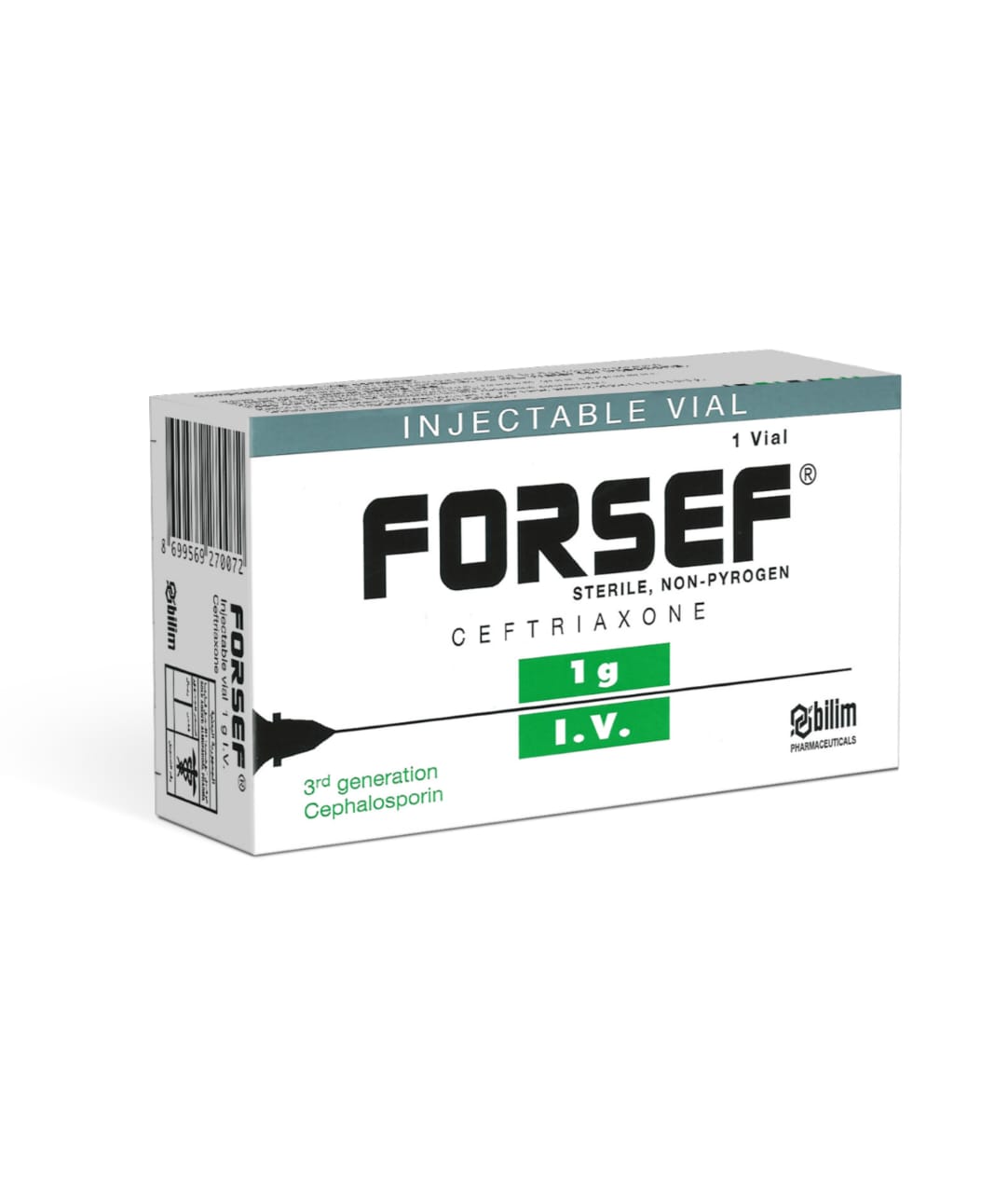
In a modern clinical hospital setting, the management of Tinea Capitis requires a precise diagnostic and therapeutic approach to differentiate fungal infections from bacterial dermatological conditions or systemic complications. While Tinea Capitis is primarily treated with systemic antifungals such as Fluconazole / فلوكونازول 150 mg or Itraconazole / إيتراكونازول 200 mg, and supplemented by topical agents like Ketoconazole Cream / كريم كيتوكونازول 2%, clinicians must remain vigilant for secondary bacterial superinfections, particularly in cases of kerion formation. Should secondary infection be suspected, targeted antibiotic therapy—such as Cefalexin / سيفالكسين 500mg or Clindamycin / كليندامايسين 300mg—may be indicated, and in severe or recalcitrant cases, wound care may involve Povidone Iodine Solution / محلول بوفيدون أيودين 100ml or Fusidic Acid Ointment / مرهم حمض الفوسيديك 2%. Furthermore, if the patient presents with systemic symptoms or signs of deep-seated infection, the clinical team may utilize Blood Cultures / مزارع الدم (خدمات رعاية عامة) to rule out bacteremia, ensuring that the treatment plan is comprehensive and appropriately integrated within our hospital’s broader diagnostic and pharmaceutical infrastructure.
Treatment & Management Options
Recommended Medications