Clinical Assessment & Protocol
Typical Presentation (HPI)
Patient presents with cyanosis unresponsive to supplemental oxygen after topical anesthetic exposure.
General Examination
Chocolate-colored blood, SpO2 gap on pulse oximetry.
Treatment Protocol
Methylene blue administration.
Patient Education
Avoid oxidizing agents and notify future providers of sensitivity.
Systemic & Specialized Examinations
EN: S1, S2 present. No murmurs. AR: صوتا القلب الأول والثاني طبيعيان. لا توجد نفخات.
EN: Lungs clear to auscultation. AR: الرئتان صافيتان عند التسمع.
EN: Abdomen soft, non-tender. AR: البطن لين ولا يوجد ألم.
EN: Alert, oriented x3. No focal deficits. AR: المريض واعي ومدرك. لا يوجد عجز عصبي بؤري.
EN: Unremarkable or not routinely indicated. AR: طبيعي أو غير مطلوب روتينياً.
EN: Unremarkable or not routinely indicated. AR: طبيعي أو غير مطلوب روتينياً.
EN: Unremarkable or not routinely indicated. AR: طبيعي أو غير مطلوب روتينياً.
EN: Unremarkable or not routinely indicated. AR: طبيعي أو غير مطلوب روتينياً.
EN: Unremarkable or not routinely indicated. AR: طبيعي أو غير مطلوب روتينياً.
Comprehensive Clinical Guide: Acquired Methemoglobinemia
1. Introduction and Overview
Acquired Methemoglobinemia (MetHb) is a potentially life-threatening hematological disorder characterized by the oxidation of the iron atom in hemoglobin from the ferrous (Fe²⁺) state to the ferric (Fe³⁺) state. While hemoglobin normally carries oxygen to tissues, the ferric form (methemoglobin) is incapable of binding oxygen. Furthermore, the presence of methemoglobin increases the affinity of the remaining ferrous heme sites for oxygen, shifting the oxyhemoglobin dissociation curve to the left, which impairs the delivery of oxygen to peripheral tissues.
Unlike congenital forms, which are typically caused by genetic deficiencies (e.g., Cytochrome b5 reductase deficiency or Hemoglobin M disease), acquired methemoglobinemia is almost exclusively secondary to exposure to oxidizing agents—be they pharmacological, environmental, or industrial. Because the condition can progress rapidly from asymptomatic cyanosis to cardiovascular collapse, it is considered a medical emergency requiring prompt recognition and intervention.
2. Etiology and Pathophysiology
The Mechanism of Oxidation
In a healthy individual, methemoglobin levels are maintained below 1% via the action of the enzyme NADH-cytochrome b5 reductase (diaphorase I). When an individual is exposed to oxidizing agents, the rate of hemoglobin oxidation exceeds the capacity of this reductase system, leading to an accumulation of methemoglobin.
Common Oxidizing Agents (Etiological Agents)
The following table categorizes the most frequent triggers for acquired methemoglobinemia:
| Category | Specific Agents |
|---|---|
| Local Anesthetics | Benzocaine, Lidocaine, Prilocaine |
| Antibiotics | Dapsone, Sulfonamides, Nitrofurantoin |
| Industrial/Chemical | Nitrites, Nitrates, Aniline dyes, Nitrobenzene |
| Analgesics | Phenacetin, Acetaminophen (rarely) |
| Others | Chlorates, Nitric oxide, Mothballs (Naphthalene) |
Pathophysiological Consequences
- Reduced Oxygen Carrying Capacity: MetHb cannot bind O₂.
- Left-Shift of Dissociation Curve: The remaining normal hemoglobin holds onto oxygen more tightly, preventing its release into the tissues (the "Haldane-like effect").
- Tissue Hypoxia: The combination of low O₂ content and impaired unloading results in profound cellular hypoxia, particularly in high-demand organs like the brain and myocardium.
3. Clinical Staging and Presentation
The clinical presentation of methemoglobinemia correlates directly with the percentage of methemoglobin in the blood. Patients often present with "chocolate-colored" blood and cyanosis that does not improve with supplemental oxygen.
| MetHb Level (%) | Clinical Manifestations |
|---|---|
| < 3% | Normal range |
| 3% – 15% | Asymptomatic; skin discoloration (slate-gray) |
| 15% – 20% | Mild cyanosis; exercise intolerance |
| 20% – 50% | Dyspnea, headache, tachycardia, fatigue, dizziness |
| 50% – 70% | Altered mental status, arrhythmias, metabolic acidosis |
| > 70% | Coma, seizures, multi-organ failure, death |
4. Diagnostic Workup and Clinical Indications
The "Saturation Gap"
A hallmark diagnostic clue is the discrepancy between the oxygen saturation measured by pulse oximetry (SpO₂) and the partial pressure of oxygen (PaO₂) measured by arterial blood gas (ABG).
* Pulse Oximetry: Often reads ~85% regardless of actual severity, as the light wavelengths used by standard pulse oximeters cannot distinguish between oxyhemoglobin and methemoglobin.
* Co-Oximetry: The gold standard. A blood gas analyzer with a co-oximeter provides the exact percentage of methemoglobin.
Differential Diagnosis
Before confirming MetHb, clinicians must rule out other causes of cyanosis and hypoxia:
1. Carbon Monoxide Poisoning: Usually presents with "cherry-red" skin, not cyanosis.
2. Sulfhemoglobinemia: A rare condition that also causes cyanosis but is resistant to treatment with methylene blue.
3. Severe Hypoxemia: Caused by pulmonary edema or pneumonia; however, in these cases, the PaO₂ will be low, whereas in MetHb, the PaO₂ remains normal.
5. Management and Therapeutic Interventions
First-Line Treatment: Methylene Blue
For symptomatic patients (typically with MetHb levels >20-30% or those with significant comorbidities), intravenous methylene blue is the treatment of choice.
* Mechanism: It acts as an electron donor for the NADPH-methemoglobin reductase pathway, effectively accelerating the reduction of Fe³⁺ back to Fe²⁺.
* Dosing: 1–2 mg/kg administered intravenously over 5 minutes.
* Caveat: Methylene blue is contraindicated in patients with Glucose-6-Phosphate Dehydrogenase (G6PD) deficiency, as it can induce severe hemolysis.
Adjunctive Therapies
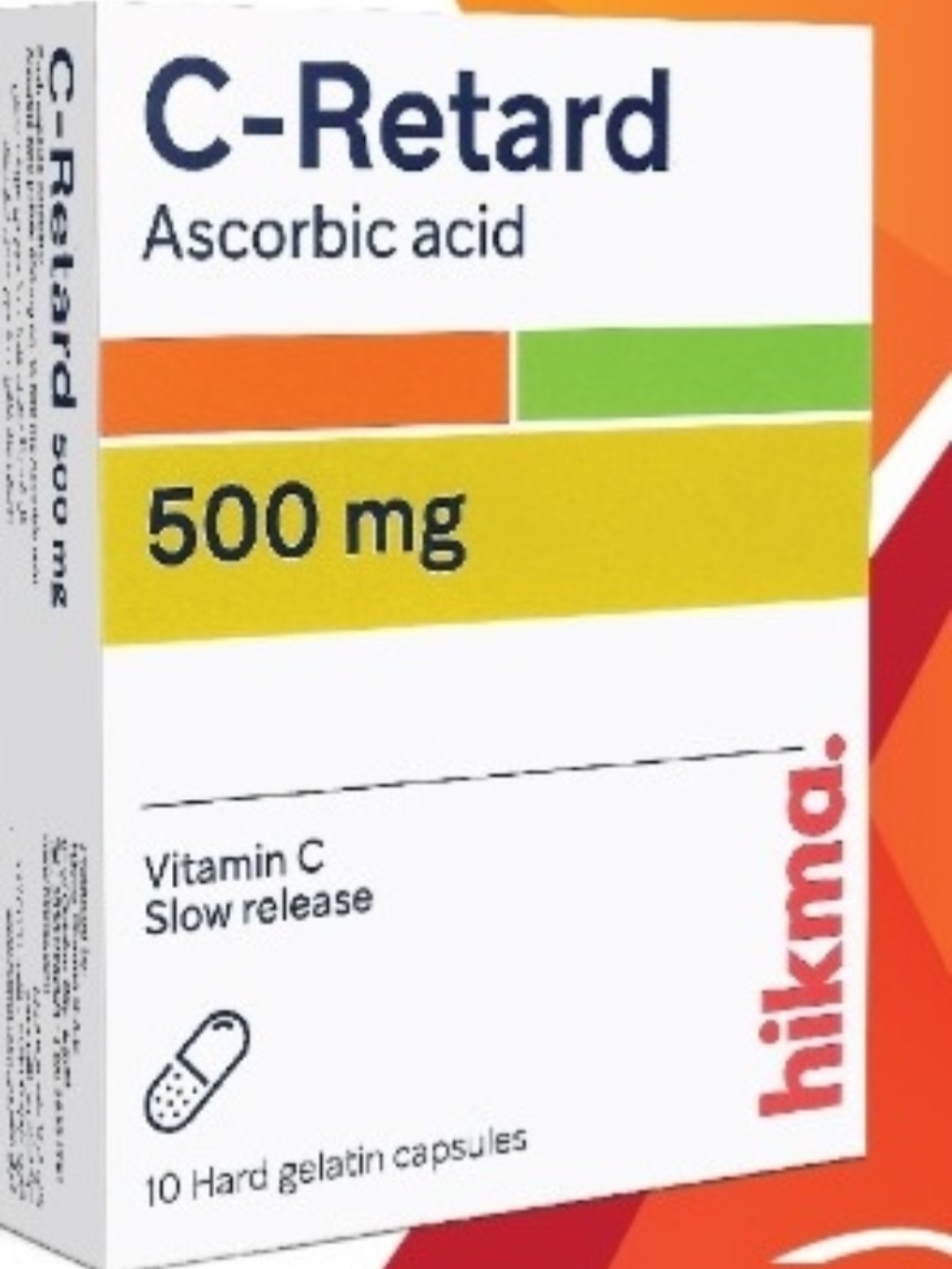
- Ascorbic Acid (Vitamin C): Useful only in mild cases or as a secondary treatment; it is a weak reducing agent.
- Exchange Transfusion: Reserved for extreme cases where methylene blue is contraindicated (e.g., G6PD deficiency) or ineffective.
- Hyperbaric Oxygen: May be considered in severe cases if other measures fail.
6. Risks, Side Effects, and Contraindications
- Paradoxical Effects: Excess methylene blue can actually cause methemoglobinemia.
- G6PD Deficiency: Always screen for this before methylene blue administration if time permits, as the drug causes oxidative stress that can trigger a hemolytic crisis in deficient patients.
- Methylene Blue Contraindications:
- Severe renal failure (often ineffective).
- Known G6PD deficiency.
- Patients taking serotonin reuptake inhibitors (risk of Serotonin Syndrome).
7. Long-term Prognosis
In cases of acquired methemoglobinemia, the prognosis is excellent if the offending agent is identified and removed. Once the methemoglobin is converted back to hemoglobin, there are typically no long-term sequelae, provided that the patient did not suffer from prolonged hypoxic brain injury during the acute phase. Patients should be counseled to avoid the causative agent for life, as recurrence is possible upon re-exposure.
8. Massive FAQ Section
Q1: Why does pulse oximetry show ~85% even when the patient is severely ill?
A: Pulse oximeters use two wavelengths of light. Methemoglobin absorbs light at both wavelengths in a way that mimics a mixture of oxyhemoglobin and deoxyhemoglobin, resulting in a "fixed" reading of approximately 85%.
Q2: How quickly does the blood color change back to red?
A: Following the administration of methylene blue, the blood typically returns to a normal bright red color within 30 to 60 minutes.
Q3: Can Acetaminophen cause methemoglobinemia?
A: Yes, though rare. It is usually associated with massive overdose or chronic, high-dose use in susceptible individuals.
Q4: Is it necessary to admit all patients with MetHb?
A: Patients with levels <20% who are asymptomatic can often be monitored in an observation unit. Symptomatic patients require ICU admission for monitoring and potential antidote administration.
Q5: What should I do if the patient has G6PD deficiency?
A: Avoid methylene blue. Use supportive care (supplemental oxygen, removal of the offending agent) and consider exchange transfusion or hyperbaric oxygen if the patient is unstable.
Q6: Does smoking contribute to MetHb levels?
A: Smoking increases baseline carboxyhemoglobin levels, but it does not directly cause methemoglobinemia. However, smokers may have less physiological reserve, making them more vulnerable to hypoxic stress.
Q7: Can I use Vitamin C instead of Methylene Blue?
A: Vitamin C is much slower and less effective. It is only appropriate for mild, asymptomatic cases where the patient is stable and the offending agent has been removed.
Q8: How do I know if the patient has "chocolate-colored" blood?
A: Clinicians often notice it during venipuncture or arterial blood gas sampling. If the blood remains dark even after exposure to air (unlike normal venous blood which turns bright red), suspect MetHb.
Q9: Can topical anesthetics like Benzocaine truly cause this?
A: Yes, Benzocaine is a frequent culprit, especially when used in excess on mucous membranes (e.g., during endoscopy or dental procedures).
Q10: Are there any long-term complications?
A: Generally, no. If the patient is resuscitated before permanent neurological or cardiac damage occurs due to hypoxia, they will make a full recovery.
9. Conclusion
Acquired methemoglobinemia is a classic medical "zebra" that clinicians must keep in their diagnostic arsenal. By maintaining a high index of suspicion in patients with unexplained cyanosis and a normal PaO₂, and by utilizing co-oximetry, the medical team can rapidly identify and treat this condition. The key to successful management lies in the swift cessation of the oxidizing agent and the judicious use of methylene blue, ensuring that the patient’s oxygen-carrying capacity is restored before irreversible tissue damage occurs.