Clinical Assessment & Protocol
Typical Presentation (HPI)
Child with developmental delay and short fourth metacarpals.
General Examination
Radiographic evidence of soft tissue calcifications and brachydactyly.
Treatment Protocol
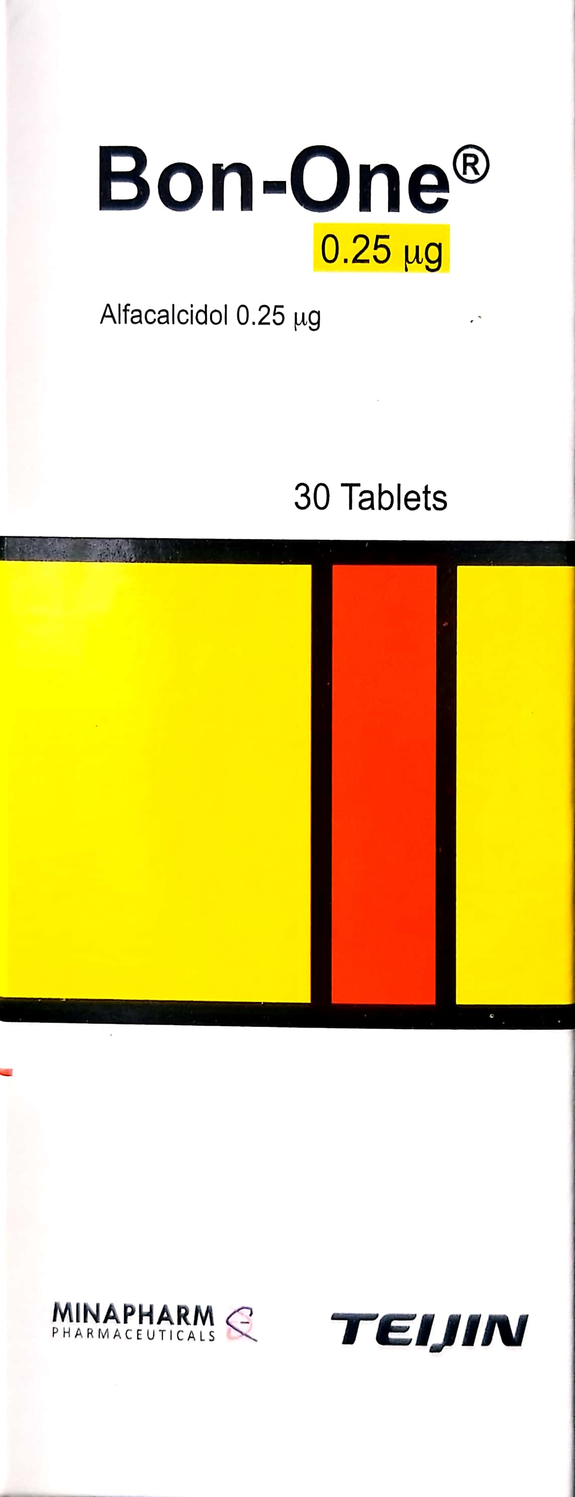
Calcium and vitamin D supplementation.
Patient Education
Regular endocrine screening is necessary.
Systemic & Specialized Examinations
EN: S1, S2 present. No murmurs. AR: صوتا القلب الأول والثاني طبيعيان. لا توجد نفخات.
EN: Lungs clear to auscultation. AR: الرئتان صافيتان عند التسمع.
EN: Abdomen soft, non-tender. AR: البطن لين ولا يوجد ألم.
EN: Alert, oriented x3. No focal deficits. AR: المريض واعي ومدرك. لا يوجد عجز عصبي بؤري.
EN: Unremarkable or not routinely indicated. AR: طبيعي أو غير مطلوب روتينياً.
EN: Unremarkable or not routinely indicated. AR: طبيعي أو غير مطلوب روتينياً.
EN: Unremarkable or not routinely indicated. AR: طبيعي أو غير مطلوب روتينياً.
EN: Unremarkable or not routinely indicated. AR: طبيعي أو غير مطلوب روتينياً.
EN: Unremarkable or not routinely indicated. AR: طبيعي أو غير مطلوب روتينياً.
Comprehensive Clinical Guide: Albright Hereditary Osteodystrophy (AHO)
Albright Hereditary Osteodystrophy (AHO) is a complex, multisystem genetic disorder characterized by a specific phenotypic presentation involving skeletal abnormalities, endocrine resistance, and often, neurodevelopmental variations. First described by Fuller Albright in 1942, the condition serves as a cornerstone study in the field of molecular endocrinology, particularly regarding G-protein signaling pathways.
This guide provides an exhaustive clinical overview intended for medical professionals, clinicians, and researchers managing patients within the AHO spectrum.
1. Clinical Definition and Overview
Albright Hereditary Osteodystrophy is a clinical phenotype resulting from mutations in the GNAS gene. It is essential to distinguish between the physical phenotype (AHO) and the biochemical presentation (Pseudohypoparathyroidism).
The AHO Phenotype
Patients presenting with the AHO phenotype exhibit:
* Short stature: Often disproportionate.
* Brachydactyly: Shortening of the metacarpals and metatarsals (typically 4th and 5th digits).
* Subcutaneous ossifications: Ectopic bone formation within the soft tissues.
* Round facies: Characteristic facial structure.
* Obesity: Often early-onset.
* Intellectual impairment: Variable severity, ranging from mild learning disabilities to moderate cognitive delay.
Diagnostic Categorization
The distinction between variants is based on the presence or absence of hormone resistance:
| Diagnosis | Phenotype (AHO) | Hormone Resistance | GNAS Imprinting |
|---|---|---|---|
| PHP-Ia | Present | Present (PTH, TSH, GHRH) | Maternal |
| PHP-Ib | Absent | Present (PTH only) | Maternal |
| PPHP | Present | Absent | Paternal |
2. Etiology and Pathophysiology
The pathophysiology of AHO is rooted in the GNAS locus on chromosome 20q13.3. This locus is subject to genomic imprinting, meaning the expression of the gene depends on whether the mutation is inherited from the mother or the father.
Molecular Mechanism
The GNAS gene encodes the alpha subunit of the stimulatory G-protein (Gsα), which is a critical mediator in the signal transduction pathway for numerous hormone receptors, including those for:
* Parathyroid Hormone (PTH)
* Thyroid Stimulating Hormone (TSH)
* Gonadotropins (FSH/LH)
* Growth Hormone Releasing Hormone (GHRH)
The Imprinting Paradox
- Maternal Inheritance (PHP-Ia): Because the Gsα protein is expressed primarily from the maternal allele in the proximal renal tubules, a maternal mutation leads to both the physical AHO phenotype and end-organ hormone resistance (the kidney fails to respond to PTH).
- Paternal Inheritance (PPHP): If the mutation is inherited from the father, the paternal allele is silenced in the renal tubules. Consequently, the maternal allele remains functional, preventing hormone resistance, yet the physical AHO phenotype persists.
3. Clinical Presentation and Staging
Clinical staging for AHO is primarily defined by the patient’s age and the progression of endocrine dysfunction.
Early Childhood (0–5 years)
- Hypocalcemia/Hyperphosphatemia: Seizures may be the first clinical indicator of PTH resistance.
- Developmental Delay: Delayed motor milestones are frequent.
- Failure to Thrive: Early growth velocity monitoring is critical.
Adolescence (10–18 years)
- Delayed Puberty: Resistance to gonadotropins may manifest as primary amenorrhea or delayed secondary sexual characteristics.
- Bone Health: Radiographic assessment of the hands often reveals the characteristic "Albright’s sign"—shortening of the 4th and 5th metacarpals.
- Obesity: BMI often accelerates during this period due to dysregulation of Gsα-mediated signaling in the hypothalamus.
Adulthood
- Metabolic Syndrome: Increased risk of insulin resistance and type 2 diabetes.
- Ectopic Calcification: Subcutaneous ossifications may cause pain or restricted range of motion in joints.
4. Key Diagnostic Tests
A robust diagnostic workup requires a multidisciplinary approach involving endocrinology, genetics, and orthopedics.
Laboratory Diagnostics
- Serum PTH, Calcium, and Phosphate: Essential to distinguish between PHP-Ia/Ib and PPHP.
- TSH and Free T4: Screening for subclinical hypothyroidism.
- Gonadotropins (FSH/LH): Assessing for hypogonadism.
- Molecular Genetic Testing: Sequencing of the GNAS gene is the gold standard for confirming the diagnosis.
Imaging Diagnostics
- Hand Radiographs: Used to identify brachydactyly (shortened metacarpals).
- DEXA Scans: Assessing bone mineral density, as patients are at risk for metabolic bone disease.
- Ultrasound/CT: Used to identify and monitor the progression of subcutaneous ossifications.
5. Risks, Complications, and Management
Management is symptomatic and centers on replacing missing hormones and monitoring systemic health.
Endocrine Management
- Hypocalcemia: Treated with active Vitamin D metabolites (calcitriol) and calcium supplementation.
- Hypothyroidism: Thyroid hormone replacement therapy (levothyroxine).
- Hypogonadism: Hormone replacement therapy as indicated by the patient’s endocrinologist.
Orthopedic Considerations
- Subcutaneous Ossification: If ossifications become symptomatic (e.g., impinging on a nerve or restricting joint movement), surgical excision may be considered, though recurrences are common.
- Bone Mineralization: High-risk patients require regular monitoring to prevent fractures.
Contraindications
- Avoid excessive PTH stimulation: Clinicians must be cautious with certain medications that may exacerbate hormonal imbalances.
- Surgical Caution: Due to the risk of abnormal bone formation, surgical intervention for ossifications should be reserved for cases where function is severely impaired.
6. Massive FAQ Section
1. Is Albright Hereditary Osteodystrophy fatal?
No, AHO is not inherently fatal. With proper management of endocrine deficiencies (particularly calcium and thyroid levels), patients can lead full, productive lives.
2. Can you have AHO without hormone resistance?
Yes. This condition is known as Pseudopseudohypoparathyroidism (PPHP). It presents with the physical features of AHO but normal biochemical markers.
3. Is there a cure for AHO?
Currently, there is no genetic cure. Treatment is focused on managing symptoms and correcting hormone deficiencies.
4. How often should a child with AHO be screened?
Annual screening for thyroid function, calcium/phosphate levels, and growth velocity is recommended.
5. Are the subcutaneous ossifications cancerous?
No, these are benign deposits of bone tissue in the skin and subcutaneous layers.
6. Does intellectual disability occur in all AHO patients?
No. While many patients experience mild to moderate cognitive impairment, there is significant variability, and some individuals have normal cognitive function.
7. Can AHO be detected prenatally?
Yes, if the specific familial mutation is known, prenatal diagnosis via amniocentesis or chorionic villus sampling is possible.
8. Is diet important for AHO patients?
Yes. Due to the propensity for obesity, a balanced diet and regular physical activity are essential to manage metabolic risks.
9. Why are the 4th and 5th fingers shorter?
This is a hallmark of the brachydactyly associated with AHO, caused by premature closure of the epiphyseal growth plates in the metacarpals.
10. Do I need to see a specialist?
Yes. AHO is a complex condition; management should be coordinated by a pediatric or adult endocrinologist, often in consultation with a clinical geneticist.
7. Long-Term Prognosis
The long-term prognosis for patients with AHO is generally positive, provided that endocrine dysfunctions are identified and managed early.
- Metabolic Health: Patients must be monitored for the development of diabetes and hypertension in adulthood.
- Quality of Life: Many individuals with AHO successfully navigate education and employment. Early intervention for learning disabilities significantly improves long-term outcomes.
- Family Planning: Given the strong genetic component, genetic counseling is vital for patients considering family planning, as the risk of passing the mutation to offspring is 50%.
Clinical Summary Table
| Feature | PHP-Ia | PHP-Ib | PPHP |
|---|---|---|---|
| AHO Phenotype | Yes | No | Yes |
| PTH Resistance | Yes | Yes | No |
| TSH/GHRH Resistance | Yes | No | No |
| Inheritance | Maternal | Maternal | Paternal |
Disclaimer: This guide is for educational purposes only and does not constitute medical advice. Diagnosis and management of Albright Hereditary Osteodystrophy should always be conducted by qualified medical professionals.