Clinical Assessment & Protocol
Typical Presentation (HPI)
Patient reports dark urine that turns black upon standing and progressive joint pain.
General Examination
Ochronosis (bluish-black pigmentation) of the sclera and ear cartilage; arthropathy of the spine.
Treatment Protocol
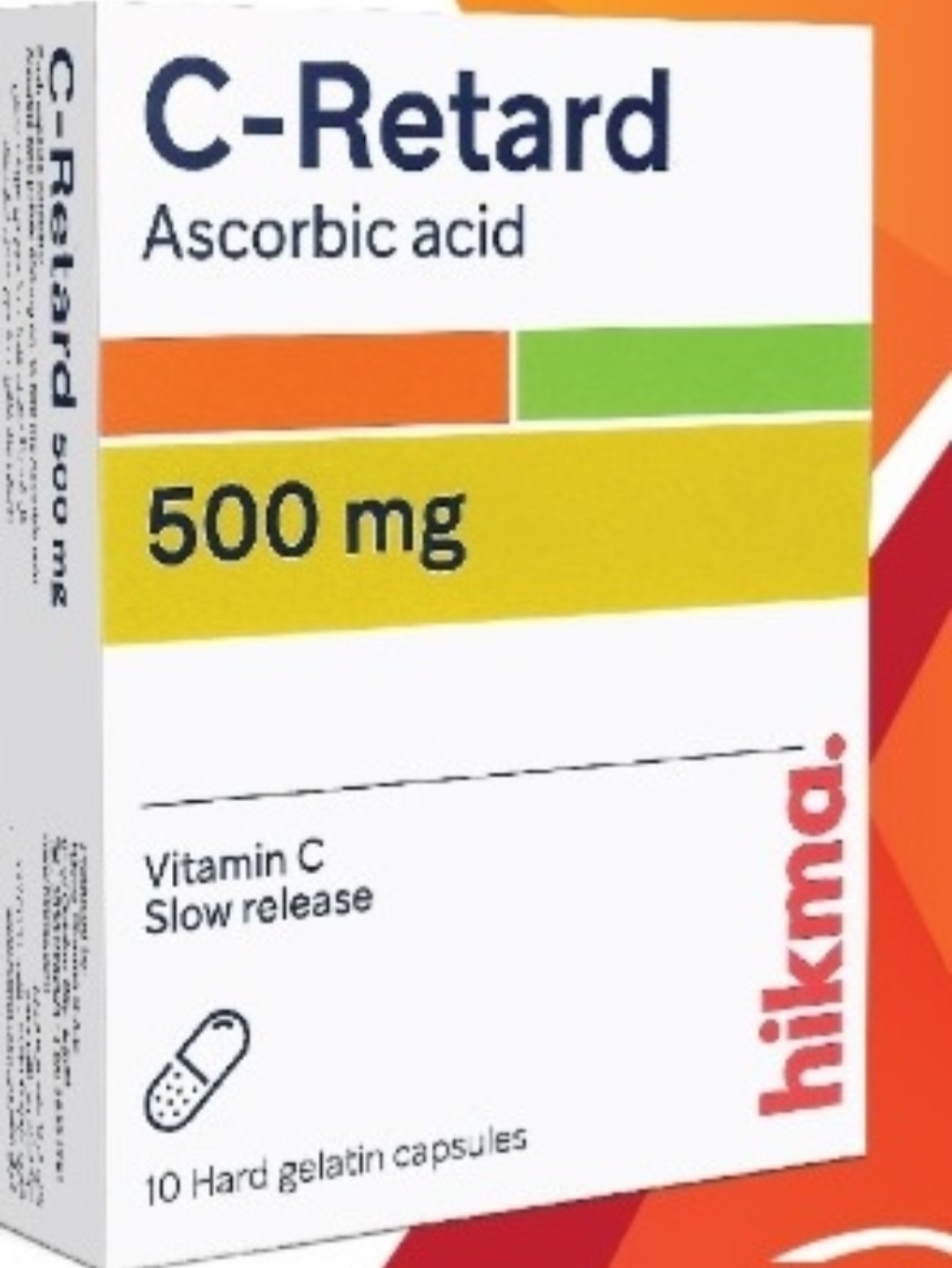
Dietary restriction of phenylalanine and tyrosine; nitisinone to inhibit homogentisic acid production.
Patient Education
Lifelong dietary management is necessary to prevent severe ochronotic arthropathy.
Systemic & Specialized Examinations
EN: S1, S2 present. No murmurs. AR: صوتا القلب الأول والثاني طبيعيان. لا توجد نفخات.
EN: Lungs clear to auscultation. AR: الرئتان صافيتان عند التسمع.
EN: Abdomen soft, non-tender. AR: البطن لين ولا يوجد ألم.
EN: Alert, oriented x3. No focal deficits. AR: المريض واعي ومدرك. لا يوجد عجز عصبي بؤري.
EN: Unremarkable or not routinely indicated. AR: طبيعي أو غير مطلوب روتينياً.
EN: Unremarkable or not routinely indicated. AR: طبيعي أو غير مطلوب روتينياً.
EN: Unremarkable or not routinely indicated. AR: طبيعي أو غير مطلوب روتينياً.
EN: Unremarkable or not routinely indicated. AR: طبيعي أو غير مطلوب روتينياً.
EN: Unremarkable or not routinely indicated. AR: طبيعي أو غير مطلوب روتينياً.
Alkaptonuria: The Comprehensive Clinical Guide
1. Comprehensive Introduction & Overview
Alkaptonuria (AKU), historically known as "black urine disease," is a rare, autosomal recessive metabolic disorder characterized by the body’s inability to fully break down the amino acids tyrosine and phenylalanine. This metabolic bottleneck results in the systemic accumulation of homogentisic acid (HGA). Over decades, this accumulation leads to a process known as ochronosis, where HGA-derived pigments deposit in connective tissues, specifically cartilage, tendons, and skin.
As an expert clinical entity, AKU represents a quintessential model of metabolic-driven degenerative joint disease. While the initial presentation may be as benign as darkened urine in infancy, the long-term clinical trajectory is one of severe, progressive, and early-onset osteoarthritis, often culminating in profound physical disability by the fifth or sixth decade of life.
Epidemiological Snapshot
- Global Prevalence: Approximately 1 in 250,000 to 1,000,000 individuals.
- Genetic Basis: Mutations in the HGD (homogentisate 1,2-dioxygenase) gene on chromosome 3q.
- Clinical Hallmark: Triad of homogentisic aciduria, ochronosis, and ochronotic arthropathy.
2. Technical Specifications & Pathophysiology
The pathophysiology of Alkaptonuria is rooted in the failure of the tyrosine catabolic pathway.
The Biochemical Mechanism
In a healthy individual, the enzyme homogentisate 1,2-dioxygenase (HGD) converts homogentisic acid into maleylacetoacetic acid. In patients with AKU, this enzyme is dysfunctional or absent. Consequently, HGA accumulates in the serum, is excreted in high concentrations in the urine, and deposits as a dark polymer in connective tissues.
| Stage | Biochemical State | Clinical Manifestation |
|---|---|---|
| Infancy | HGA excretion | Darkening urine upon oxidation |
| Adolescence | Tissue deposition | Scleral/aural pigmentation |
| Adulthood | Ochronosis | Progressive joint stiffness/pain |
| Geriatrics | Systemic failure | Severe arthropathy, cardiac valve calcification |
The Ochronotic Process
The term "ochronosis" refers to the blue-black discoloration of connective tissues. HGA oxidizes into benzoquinone acetic acid (BQA), which polymerizes to form melanin-like pigments. These pigments bind to collagen fibers, rendering cartilage brittle, less resilient to mechanical stress, and prone to fragmentation.
3. Clinical Indications & Usage: The Diagnostic Pathway
Diagnosis requires a high index of clinical suspicion, as many patients remain asymptomatic until their 30s or 40s.
Standard Presentation
- Urine Discoloration: Parents may notice diapers turning black. However, this is often missed if the urine is not left to stand for several hours to allow for oxidation.
- Ocular/Dermal Signs: The earliest visible signs include blue-gray pigmentation of the sclera (Osler’s sign) and the pinna of the ear.
- Musculoskeletal Presentation:
- Spinal involvement: Early-onset intervertebral disc calcification, leading to "bamboo spine" appearance (distinct from Ankylosing Spondylitis).
- Peripheral joints: Chronic pain and stiffness in large weight-bearing joints (knees, hips, shoulders).
Key Diagnostic Tests
- Urine Analysis (Gas Chromatography-Mass Spectrometry): The gold standard for measuring HGA levels.
- Alkaline Test: Adding sodium hydroxide to urine causes it to turn black almost instantly.
- Radiographic Imaging:
- Spine: Dense calcification of intervertebral discs.
- Joints: Narrowing of joint spaces, subchondral sclerosis, and presence of intra-articular loose bodies (osteochondral fragments).
- Genetic Testing: Sequencing of the HGD gene confirms the diagnosis by identifying biallelic mutations.
4. Risks, Side Effects, and Long-Term Prognosis
The prognosis of AKU is dictated by the severity of ochronotic arthropathy and systemic complications.
Complications and Risks
- Cardiac Involvement: The most significant risk factor for mortality. HGA deposits in heart valves (mitral and aortic), leading to calcification, stenosis, and regurgitation.
- Renal Stones: Approximately 50% of patients develop renal calculi composed of calcium oxalate.
- Prostate Stones: Common in male patients due to the concentration of HGA in prostatic secretions.
- Tendon Rupture: Achillean and other major tendons become brittle and are prone to spontaneous rupture.
Management Strategies
While there is no "cure," modern management focuses on mitigating HGA accumulation:
1. Nitisinone Therapy: An FDA-approved (in some regions) herbicide that inhibits the enzyme 4-hydroxyphenylpyruvate dioxygenase, effectively blocking HGA production upstream.
2. Dietary Modification: Low-protein diets (limiting phenylalanine and tyrosine) are often recommended, though their efficacy in adult patients is debated.
3. Orthopedic Intervention: Total joint arthroplasty (TJA) is frequently required for hips and knees. Surgeons must be aware of the brittle bone quality and potential for prolonged healing.
5. Frequently Asked Questions (FAQ)
1. Is Alkaptonuria fatal?
AKU itself is not typically fatal, but the associated cardiac complications (valvular heart disease) and potential for severe physical disability significantly impact life expectancy and quality of life.
2. Can I pass this to my children?
Yes. As an autosomal recessive disorder, if both parents are carriers, there is a 25% chance of each child having the condition. Genetic counseling is highly recommended.
3. Does the urine color change immediately?
No. Fresh urine is usually normal in color. It turns dark upon exposure to air (oxidation) or when exposed to alkaline solutions.
4. Is there a specific diet for AKU?
A low-protein diet is often suggested to reduce the intake of tyrosine and phenylalanine. However, it must be carefully managed by a dietician to avoid malnutrition.
5. Why does it take so long to diagnose?
Because the symptoms of joint pain are non-specific and mimic common osteoarthritis, clinicians often overlook the possibility of a metabolic cause until the disease is advanced.
6. What is the difference between AKU and Ankylosing Spondylitis?
While both cause spinal stiffness, AKU is a metabolic disease with disc calcification, whereas AS is an inflammatory autoimmune disease characterized by syndesmophytes and fusion.
7. Does Nitisinone reverse the damage?
Nitisinone stops the production of HGA, preventing further accumulation. It cannot reverse existing pigment deposits or repair damaged cartilage, which is why early initiation is vital.
8. Are there any skin changes?
Yes, "ochronosis" can cause darkening of the skin in areas with high sweat gland density, such as the axillae or the bridge of the nose.
9. What should I avoid if I have AKU?
Avoid high-protein supplements, heavy contact sports that stress the joints, and any unnecessary physical trauma to the tendons.
10. Can I have a total joint replacement?
Yes, and it is often necessary. However, surgeons must be alerted to the condition as the bone quality may be compromised by pigment deposition.
6. Clinical Summary & Expert Recommendations
Alkaptonuria remains a challenging clinical diagnosis due to its rarity and the long latency period between biochemical onset and clinical symptomatic presentation. From an orthopedic perspective, the management of the AKU patient requires a multidisciplinary approach involving:
- Metabolic Specialists: To monitor HGA levels and manage Nitisinone dosing.
- Cardiologists: Annual echocardiograms are mandatory for patients over 40 to monitor for valvular calcification.
- Orthopedic Surgeons: To manage the inevitable arthropathy with specialized surgical techniques for brittle connective tissue.
Differential Diagnosis Table
| Condition | Primary Distinguishing Factor |
|---|---|
| Osteoarthritis (Primary) | Age of onset, absence of systemic/metabolic markers. |
| Ankylosing Spondylitis | Inflammatory markers (CRP/ESR), HLA-B27 positivity. |
| Hemochromatosis | Iron overload, specific arthropathy in MCP joints. |
| Wilson’s Disease | Copper metabolism, hepatic and neurological involvement. |
In conclusion, early recognition of the hallmark signs—dark urine and scleral pigmentation—is the most effective tool in the clinical arsenal. By transitioning from reactive treatment to proactive metabolic management, the clinical prognosis for individuals with Alkaptonuria can be significantly improved, shifting the narrative from inevitable disability to managed, active living.
Medical Disclaimer: This guide is for educational purposes only and does not constitute medical advice, diagnosis, or treatment. Always seek the advice of your physician or other qualified health provider with any questions regarding a medical condition.