Clinical Assessment & Protocol
Typical Presentation (HPI)
EN: Patient presents with clinical signs of burn wound infection, including increased pain, purulent exudate, foul odor, and periwound erythema. Onset of symptoms noted [Time/Days] post-injury. Associated with [fever/chills/malaise]. No prior antibiotic therapy reported. AR: يراجع المريض بأعراض سريرية تدل على عدوى في حرق، تشمل زيادة في الألم، إفرازات قيحية، رائحة كريهة، واحمرار حول الجرح. بدأت الأعراض منذ [الوقت/الأيام] بعد الإصابة. ترافق ذلك مع [حمى/قشعريرة/توعك]. لا يوجد تاريخ لاستخدام مضادات حيوية سابقة.
General Examination
EN: Physical examination reveals [percentage]% TBSA burn site with localized cellulitis, increased warmth, and purulent discharge. Wound bed shows [slough/eschar/granulation tissue]. Periwound edema present. Systemic signs: [stable/tachycardic/febrile]. Palpable regional lymphadenopathy noted. AR: يكشف الفحص السريري عن موقع حرق بنسبة [النسبة]% من مساحة سطح الجسم مع التهاب خلوي موضعي، زيادة في الحرارة، وإفرازات قيحية. يظهر قاع الجرح [نخر/قشور/نسيج حبيبي]. يوجد وذمة حول الجرح. العلامات الجهازية: [مستقرة/تسرع قلب/حمى]. لوحظ وجود ضخامة عقد لمفاوية إقليمية.
Treatment Protocol
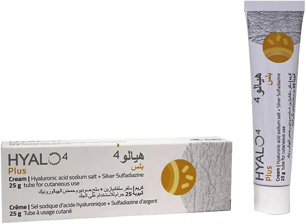
EN: Immediate wound debridement performed. Wound culture and sensitivity obtained. Initiated empiric IV antibiotics [Drug Name/Dosage]. Topical antimicrobial therapy [e.g., Silver Sulfadiazine/Mafenide Acetate] applied. Daily dressing changes with sterile technique. Monitor for systemic sepsis. AR: تم إجراء تنضير فوري للجرح. تم أخذ مسحة من الجرح للزرع والتحسس. بدأ العلاج بالمضادات الحيوية الوريدية التجريبية [اسم الدواء/الجرعة]. تم تطبيق علاج مضاد للميكروبات موضعي [مثل: سلفاديازين الفضة/أسيتات المافينيد]. تغيير الضمادات يومياً باتباع تقنية معقمة. المراقبة المستمرة للكشف عن أي علامات للإنتان الجهازي.
Patient Education
EN: Maintain strict hygiene during dressing changes. Monitor for worsening symptoms: spreading redness, fever >38°C, or increased pain. Keep the burn site clean and dry unless instructed otherwise. Adhere strictly to the prescribed antibiotic regimen. Follow-up appointment scheduled for [Date/Time]. AR: حافظ على نظافة صارمة أثناء تغيير الضمادات. راقب الأعراض التي تشير إلى تفاقم الحالة: انتشار الاحمرار، حمى > 38 درجة مئوية، أو زيادة في الألم. حافظ على موقع الحرق نظيفاً وجافاً ما لم يتم توجيهك بخلاف ذلك. التزم بدقة بنظام المضادات الحيوية الموصوف. موعد المراجعة القادم في [التاريخ/الوقت].
Systemic & Specialized Examinations
EN: Unremarkable. Systemic examination is not the primary focus for this reconstructive or aesthetic presentation. AR: طبيعي. الفحص الجهازي ليس التركيز الأساسي لهذه الحالة التجميلية أو الترميمية.
EN: Unremarkable. Systemic examination is not the primary focus for this reconstructive or aesthetic presentation. AR: طبيعي. الفحص الجهازي ليس التركيز الأساسي لهذه الحالة التجميلية أو الترميمية.
EN: Unremarkable. Systemic examination is not the primary focus for this reconstructive or aesthetic presentation. AR: طبيعي. الفحص الجهازي ليس التركيز الأساسي لهذه الحالة التجميلية أو الترميمية.
EN: Unremarkable. Systemic examination is not the primary focus for this reconstructive or aesthetic presentation. AR: طبيعي. الفحص الجهازي ليس التركيز الأساسي لهذه الحالة التجميلية أو الترميمية.
EN: Focused assessment of the affected anatomical sub-unit (skin, soft tissue, bone). Findings are consistent with Burn Wound Infection. Pre-operative photography and planning performed. AR: فحص موجه للوحدة التشريحية المصابة (الجلد، الأنسجة الرخوة، العظام). النتائج تتوافق مع Burn Wound Infection. تم إجراء التصوير والتخطيط قبل الجراحة.
EN: Unremarkable. Systemic examination is not the primary focus for this reconstructive or aesthetic presentation. AR: طبيعي. الفحص الجهازي ليس التركيز الأساسي لهذه الحالة التجميلية أو الترميمية.
EN: Unremarkable. Systemic examination is not the primary focus for this reconstructive or aesthetic presentation. AR: طبيعي. الفحص الجهازي ليس التركيز الأساسي لهذه الحالة التجميلية أو الترميمية.
EN: Unremarkable. Systemic examination is not the primary focus for this reconstructive or aesthetic presentation. AR: طبيعي. الفحص الجهازي ليس التركيز الأساسي لهذه الحالة التجميلية أو الترميمية.
EN: Unremarkable. Systemic examination is not the primary focus for this reconstructive or aesthetic presentation. AR: طبيعي. الفحص الجهازي ليس التركيز الأساسي لهذه الحالة التجميلية أو الترميمية.
Orthopedic & Trauma Assessments
EN: Unremarkable. Systemic examination is not the primary focus for this reconstructive or aesthetic presentation. AR: طبيعي. الفحص الجهازي ليس التركيز الأساسي لهذه الحالة التجميلية أو الترميمية.
EN: Unremarkable. Systemic examination is not the primary focus for this reconstructive or aesthetic presentation. AR: طبيعي. الفحص الجهازي ليس التركيز الأساسي لهذه الحالة التجميلية أو الترميمية.
EN: Unremarkable. Systemic examination is not the primary focus for this reconstructive or aesthetic presentation. AR: طبيعي. الفحص الجهازي ليس التركيز الأساسي لهذه الحالة التجميلية أو الترميمية.
EN: Unremarkable. Systemic examination is not the primary focus for this reconstructive or aesthetic presentation. AR: طبيعي. الفحص الجهازي ليس التركيز الأساسي لهذه الحالة التجميلية أو الترميمية.
EN: Unremarkable. Systemic examination is not the primary focus for this reconstructive or aesthetic presentation. AR: طبيعي. الفحص الجهازي ليس التركيز الأساسي لهذه الحالة التجميلية أو الترميمية.
EN: Unremarkable. Systemic examination is not the primary focus for this reconstructive or aesthetic presentation. AR: طبيعي. الفحص الجهازي ليس التركيز الأساسي لهذه الحالة التجميلية أو الترميمية.
EN: Unremarkable. Systemic examination is not the primary focus for this reconstructive or aesthetic presentation. AR: طبيعي. الفحص الجهازي ليس التركيز الأساسي لهذه الحالة التجميلية أو الترميمية.
EN: Unremarkable. Systemic examination is not the primary focus for this reconstructive or aesthetic presentation. AR: طبيعي. الفحص الجهازي ليس التركيز الأساسي لهذه الحالة التجميلية أو الترميمية.
EN: Unremarkable. Systemic examination is not the primary focus for this reconstructive or aesthetic presentation. AR: طبيعي. الفحص الجهازي ليس التركيز الأساسي لهذه الحالة التجميلية أو الترميمية.
Burn Wound Infection: A Comprehensive Medical SEO Guide
Burns are a significant cause of morbidity and mortality worldwide. While the initial thermal injury is critical, the subsequent development of a burn wound infection represents a major complication that can drastically alter the patient's prognosis. This guide, tailored for patients and their families, provides an in-depth understanding of burn wound infections from the perspective of a reconstructive and plastic surgery specialist. We will explore the underlying causes, the intricate biological processes involved, how to recognize the signs and symptoms, the diagnostic approaches, and the current standards of care for treatment and long-term management.
1. Executive Overview: Understanding Burn Wound Infection
A burn wound infection is defined as the invasion and multiplication of microorganisms within a burn wound that leads to a host immune response and potential systemic complications. The compromised skin barrier in a burn patient creates a prime environment for bacterial, fungal, and occasionally viral pathogens to colonize and proliferate. This colonization can progress to invasive infection, where microorganisms breach the wound bed and enter the bloodstream or deeper tissues, leading to sepsis, multi-organ failure, and increased mortality.
The severity of a burn (depth, size, and location) significantly influences the risk of infection. Deep partial-thickness and full-thickness burns are particularly vulnerable due to the destruction of the protective epidermis and dermis. Burn wound infections are a leading cause of death in burn patients, even with advancements in critical care and antimicrobial therapies. Early recognition, aggressive management, and a multidisciplinary approach involving burn surgeons, infectious disease specialists, and wound care nurses are paramount for optimal outcomes.
Key Takeaways:
- Definition: Microbial invasion and multiplication in a burn wound.
- Significance: A leading cause of mortality and morbidity in burn patients.
- Vulnerability: Deep and large burns are at highest risk.
- Goal: Early detection, prompt treatment, and prevention.
2. Detailed Pathophysiology, Etiology, and Risk Factors
Understanding the biological cascade of burn wound infection is crucial to appreciating its severity and the rationale behind treatment strategies.
Pathophysiology: A Breach in Defense
The skin is our body's primary defense against external pathogens. A burn injury profoundly disrupts this barrier in several ways:
- Loss of Integumentary Barrier: The destruction of the epidermis and dermis removes the physical barrier that prevents microbial entry.
- Eschar Formation: In deep burns, a dry, devitalized tissue (eschar) forms. This eschar is a nutrient-rich, avascular environment that can harbor bacteria and hinder the delivery of topical antimicrobials and immune cells.
- Edema: Burn-induced inflammation causes significant edema (swelling) in the burn area and surrounding tissues. This edema can impair local blood flow, reducing the delivery of oxygen, nutrients, and immune cells to the wound, thereby compromising the local immune response.
- Compromised Immune System: The systemic inflammatory response to a major burn can paradoxically lead to immunosuppression, making the patient more susceptible to infections. Cytokine dysregulation and impaired neutrophil function contribute to this.
- Moisture and Nutrient Rich Environment: Burn exudate provides a moist, protein-rich environment ideal for bacterial growth.
Once pathogens colonize the wound, they can proliferate. If the host immune system cannot contain this proliferation, the microorganisms can invade the viable tissue beneath the eschar or wound bed. This invasive phase is characterized by local tissue destruction and can lead to the release of toxins and inflammatory mediators that further damage host tissues. The infection can then spread hematogenously (through the bloodstream) to distant organs, leading to sepsis, a life-threatening condition.
Etiology: The Culprits of Infection
The microorganisms responsible for burn wound infections are diverse and often depend on the environment and the patient's immune status.
-
Common Bacterial Pathogens:
- Staphylococcus aureus: A ubiquitous bacterium, often found on the skin. Methicillin-resistant Staphylococcus aureus (MRSA) is a significant concern.
- Pseudomonas aeruginosa: A gram-negative bacterium frequently found in the environment (soil, water). It thrives in moist wounds and is notorious for its rapid proliferation, production of toxins, and development of antibiotic resistance. It often produces a characteristic blue-green pus and a fruity odor.
- Streptococcus pyogenes (Group A Streptococcus): Can cause rapid and severe soft tissue infections, including cellulitis and necrotizing fasciitis.
- Enterococcus species: Increasingly recognized as significant pathogens, especially in patients with prolonged hospital stays or prior antibiotic exposure.
- Acinetobacter baumannii: A gram-negative bacterium that can cause severe, difficult-to-treat infections, particularly in intensive care settings.
-
Fungal Pathogens:
- Candida species: Common commensals, but can overgrow in burn wounds, especially with prolonged antibiotic use or immunosuppression.
- Aspergillus species: Can cause invasive infections, particularly in severely immunocompromised patients.
-
Viral Pathogens: Less common as primary burn wound invaders but can complicate the healing process, especially in immunocompromised individuals.
Risk Factors: Who is Most Vulnerable?
Several factors increase a patient's susceptibility to burn wound infections:
- Burn Size: Larger burn surface area correlates with higher infection risk due to a greater area of compromised skin and a more significant systemic inflammatory response.
- Burn Depth: Deep partial-thickness and full-thickness burns are at higher risk than superficial burns.
- Location of Burn: Burns on the extremities, perineum, and trunk are more prone to contamination.
- Delayed Wound Excision: Delaying the surgical removal of necrotic eschar allows more time for bacterial colonization and proliferation.
- Prolonged Hospitalization: Extended stays increase exposure to hospital-acquired pathogens.
- Immunosuppression: Conditions or treatments that suppress the immune system (e.g., chemotherapy, HIV, corticosteroid use) significantly increase risk.
- Malnutrition: Poor nutritional status impairs immune function and wound healing.
- Presence of Foreign Bodies: Devices like urinary catheters or central venous lines can serve as portals of entry for infection.
- Inadequate Wound Care: Poor hygiene or insufficient application of topical antimicrobials.
- Age: Very young children and the elderly have less robust immune systems.
3. Signs, Symptoms, and Clinical Presentation
Recognizing the signs and symptoms of burn wound infection is critical for early intervention. While some signs are local to the wound, others can indicate systemic spread.
Local Signs and Symptoms: What to Look For in the Wound
- Increased Pain: While some discomfort is expected after a burn, a sudden or disproportionate increase in pain, especially in areas that were previously less painful, can be a sign of infection.
- Wound Discoloration: Changes in the color of the wound bed or exudate. A healthy wound bed is typically pink or red. Signs of infection can include a pale or grayish appearance, or the presence of greenish-yellow pus.
- Malodor: A foul or unusual smell emanating from the wound is a common indicator of bacterial infection, particularly Pseudomonas aeruginosa.
- Increased Exudate: A significant increase in the amount of wound drainage, which may also change in color or consistency (e.g., becoming purulent).
- Edema and Erythema: Increased swelling (edema) and redness (erythema) spreading beyond the immediate burn margin.
- Eschar Changes: The eschar may become softened, friable, or develop dark spots (necrotic areas) within it. In some cases, tunneling under the eschar can occur.
- Delayed Granulation: The formation of healthy granulation tissue (new connective tissue and blood vessels) is essential for wound healing. Infection can impede or halt this process.
- Sloughing of Grafted Skin: If skin grafts have been applied, signs of infection can lead to graft failure and sloughing.
Systemic Signs and Symptoms: When Infection Spreads
Systemic signs indicate that the infection is no longer localized and is affecting the entire body. These are medical emergencies.
- Fever or Hypothermia: An elevated body temperature (fever, >38.3°C or 101°F) is a classic sign of infection. Conversely, hypothermia (low body temperature, <36°C or 96.8°F) can also occur in severe sepsis.
- Tachycardia: An abnormally rapid heart rate (>100 beats per minute in adults).
- Tachypnea: Abnormally rapid breathing (>20 breaths per minute in adults).
- Hypotension: Low blood pressure, which can lead to dizziness, lightheadedness, or fainting.
- Altered Mental Status: Confusion, lethargy, disorientation, or decreased responsiveness.
- Decreased Urine Output: Reduced kidney function can lead to a significant decrease in urine production.
- Leukocytosis or Leukopenia: Elevated white blood cell count (leukocytosis) or a dangerously low white blood cell count (leukopenia) in blood tests.
- Metabolic Acidosis: A buildup of acid in the body, detectable through blood gas analysis.
It is crucial to report any of these signs and symptoms to your medical team immediately.
4. Standard Diagnostic Evaluation & Workup
Diagnosing burn wound infection involves a combination of clinical assessment, laboratory tests, and sometimes imaging. The goal is to confirm the presence of infection, identify the causative organism, and assess its extent.
Clinical Diagnosis
The initial suspicion of infection is largely based on the clinical presentation described above. A thorough physical examination of the burn wound and assessment for systemic signs are the first steps.
Laboratory Assays
Several laboratory tests are vital in diagnosing and monitoring burn wound infections:
-
Wound Cultures:
- Quantitative Biopsy Culture: This is considered the gold standard for diagnosing burn wound infection. A small piece of viable tissue from the wound bed is surgically removed (biopsied) and sent to the laboratory for bacterial and fungal culture. A bacterial count of ≥10^5 organisms per gram of tissue is generally considered diagnostic of invasive burn wound infection.
- Swab Cultures: While less definitive than biopsies, superficial swab cultures can help identify the predominant organisms present on the wound surface. They are often used for initial screening or when biopsy is not immediately feasible.
-
Blood Cultures: If systemic infection (sepsis) is suspected, blood cultures are essential to identify microorganisms in the bloodstream and determine antibiotic sensitivities.
-
Complete Blood Count (CBC) with Differential: This test assesses the number of white blood cells (WBCs), red blood cells, and platelets. An elevated WBC count (leukocytosis) or a depressed WBC count (leukopenia) can indicate infection or an overwhelming inflammatory response.
-
Inflammatory Markers:
- C-Reactive Protein (CRP): A protein produced by the liver in response to inflammation. Elevated CRP levels are indicative of an inflammatory process, including infection.
- Procalcitonin: A biomarker that is more specific for bacterial infections than CRP. Elevated levels can help distinguish between bacterial and non-bacterial causes of inflammation.
-
Renal and Liver Function Tests: Blood tests to assess kidney (e.g., BUN, creatinine) and liver function (e.g., AST, ALT, bilirubin) are important for monitoring organ damage due to sepsis.
-
Arterial Blood Gases (ABGs): Used to assess oxygenation, ventilation, and acid-base balance, particularly in patients with suspected sepsis or respiratory compromise.
Imaging Studies
Imaging plays a supportive role in evaluating the extent of infection or complications.
- X-rays: Can be useful for identifying deep tissue gas, which may indicate anaerobic infection or gas gangrene, or for detecting foreign bodies.
- Ultrasound: Can help visualize soft tissue collections such as abscesses or hematomas, and assess local blood flow.
- Computed Tomography (CT) Scan: Useful for delineating deeper infections, abscesses, or osteomyelitis (bone infection) in adjacent structures.
- Magnetic Resonance Imaging (MRI): Provides excellent soft tissue detail and is often used to assess the extent of deep tissue infection or involvement of underlying structures.
5. Therapeutic Interventions: A Multifaceted Approach
The treatment of burn wound infection is aggressive and often requires a multidisciplinary approach. It typically involves a combination of pharmacotherapy, surgical debridement, and supportive care.
Pharmacotherapy (Antibiotics and Antifungals)
-
Empirical Antibiotic Therapy: Treatment is usually initiated empirically (based on the most likely pathogens) as soon as infection is suspected, pending culture results. The choice of antibiotics depends on local resistance patterns, the patient's history, and the severity of the infection.
- Topical Antimicrobials: These are applied directly to the burn wound to control microbial colonization and prevent invasion. Common agents include:
- Silver sulfadiazine (SSD): A broad-spectrum agent, though resistance can develop.
- Mafenide acetate: Effective against a wide range of bacteria, including Pseudomonas, but can cause pain and metabolic acidosis.
- Silver-impregnated dressings: Offer sustained release of silver ions.
- Bacitracin/Neomycin/Polymyxin B: Often used for superficial burns.
- Systemic Antibiotics: If the infection is invasive or there are signs of systemic spread, intravenous (IV) antibiotics are administered. The selection is guided by culture and sensitivity results once available. The goal is to achieve adequate drug concentrations at the wound site and in the bloodstream.
- Topical Antimicrobials: These are applied directly to the burn wound to control microbial colonization and prevent invasion. Common agents include:
-
Antifungal Therapy: If fungal infection is identified, systemic or topical antifungal medications will be prescribed.
Surgical Interventions
Surgical management is a cornerstone of burn wound infection treatment.
-
Debridement:
- Tangential Excision: This involves the surgical removal of thin layers of devitalized (necrotic) tissue down to viable tissue. It is performed meticulously to remove infected or non-viable tissue, promote healing, and prepare the wound bed for grafting.
- Escharotomy and Fasciotomy: For circumferential burns that constrict blood flow or breathing, incisions (escharotomy or fasciotomy) are made through the eschar to relieve pressure and restore circulation.
-
Wound Excision and Grafting: Once the infection is controlled and the wound bed is healthy, definitive surgical closure with autografts (skin from the patient's own body) or skin substitutes is performed to restore the skin barrier.
-
Incision and Drainage of Abscesses: If localized pus collections (abscesses) are present, they must be surgically drained.
Lifestyle and Supportive Care
- Nutritional Support: Adequate nutrition is vital for immune function and wound healing. This often involves high-protein, high-calorie diets, sometimes supplemented with enteral or parenteral nutrition.
- Pain Management: Effective pain control is essential for patient comfort and to facilitate wound care procedures.
- Fluid and Electrolyte Balance: Burn patients are at risk for dehydration and electrolyte imbalances due to fluid loss and the inflammatory response. Intravenous fluids are crucial for maintaining hemodynamic stability.
- Wound Care Protocols: Strict adherence to sterile dressing changes, wound cleansing, and infection control measures is paramount.
- Isolation Precautions: Patients with highly contagious or multidrug-resistant organisms may require isolation to prevent transmission.
6. Frequently Asked Questions (FAQ)
Here are answers to some of the most common questions patients have about burn wound infections.
Q1: How do I know if my burn is infected?
A1: Signs of infection include increased pain, foul odor, changes in wound color (e.g., green or yellow pus), increased wound drainage, swelling, and redness spreading beyond the burn. Systemic signs like fever, chills, rapid heart rate, or confusion are also critical indicators and require immediate medical attention.
Q2: What is the most common cause of burn wound infections?
A2: The most common culprits are bacteria, particularly Staphylococcus aureus and Pseudomonas aeruginosa. Fungi like Candida can also cause infections, especially in patients on prolonged antibiotics.
Q3: Is a burn wound infection always serious?
A3: Yes, any burn wound infection should be taken very seriously. While some infections can be localized and treated effectively with topical agents, they have the potential to spread to the bloodstream (sepsis), leading to life-threatening complications and organ failure.
Q4: What is the "gold standard" test for diagnosing burn wound infection?
A4: The gold standard is a quantitative wound biopsy culture. This involves surgically taking a small sample of viable tissue from the burn wound bed and sending it to the lab for precise bacterial counts. A count of 10^5 or more organisms per gram of tissue is generally considered diagnostic of invasive infection.
Q5: How are burn wound infections treated?
A5: Treatment typically involves a combination of strategies:
* Topical antimicrobials: Applied directly to the wound.
* Systemic antibiotics: Given intravenously if the infection is severe or spreading.
* Surgical debridement: Removal of infected or dead tissue.
* Wound cleaning and dressing changes: Performed meticulously and regularly.
* Nutritional support and pain management.
Q6: Can burn wound infections be prevented?
A6: Prevention is a critical aspect of burn care. Strategies include:
* Prompt and thorough wound cleaning.
* Early surgical removal of dead tissue (eschar).
* Appropriate and timely application of topical antimicrobial agents.
* Strict adherence to sterile techniques during wound care.
* Good hand hygiene by healthcare providers and visitors.
* Maintaining adequate nutrition and immune function.
Q7: How long does it take to recover from a burn wound infection?
A7: The recovery time varies significantly depending on the severity of the burn, the extent of the infection, the type of organism involved, and the patient's overall health. It can range from weeks to months, often requiring multiple surgeries and extensive rehabilitation.
Q8: What are the long-term consequences of a burn wound infection?
A8: Burn wound infections can lead to delayed healing, increased scarring, contractures (tightening of skin and underlying tissues), chronic pain, and functional impairment. In severe cases, they can result in disfigurement, amputation, or even death.
Q9: When should I go to the emergency room for a burn?
A9: You should seek immediate emergency medical care for any burn that is:
* Larger than your palm.
* Deep (causes blistering or appears white/leathery).
* Caused by chemicals or electricity.
* Located on the face, hands, feet, genitals, or over a major joint.
* If you suspect infection (as described in Q1).
Q10: What is the role of plastic and reconstructive surgery in managing burn wound infections?
A10: Plastic and reconstructive surgeons are central to managing burn wounds and their complications, including infections. They are experts in:
* Assessing burn severity.
* Performing surgical debridement of infected and necrotic tissue.
* Controlling bleeding and managing fluid shifts.
* Selecting and applying appropriate antimicrobial strategies.
* Performing skin grafting and reconstructive procedures to close the wound and restore function and appearance.
* Managing long-term complications such as scarring and contractures.
This comprehensive guide aims to empower patients with knowledge about burn wound infections. Always consult with your medical team for personalized advice and treatment plans.