Clinical Assessment & Protocol
Typical Presentation (HPI)
Painless, slowly enlarging skin nodule leading to a large ulcer with undermined edges.
General Examination
Large, deep ulcer with necrotic base and undermined edges.
Treatment Protocol
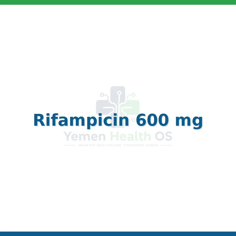
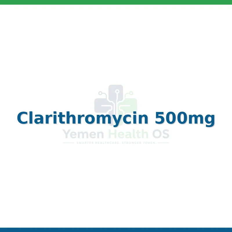
Combination of Rifampicin and Clarithromycin.
Systemic & Specialized Examinations
EN: S1, S2 present. No murmurs. AR: صوتا القلب الأول والثاني طبيعيان. لا توجد نفخات.
EN: Lungs clear to auscultation. AR: الرئتان صافيتان عند التسمع.
EN: Abdomen soft, non-tender. AR: البطن لين ولا يوجد ألم.
EN: Alert, oriented x3. No focal deficits. AR: المريض واعي ومدرك. لا يوجد عجز عصبي بؤري.
EN: Unremarkable or not routinely indicated. AR: طبيعي أو غير مطلوب روتينياً.
EN: Unremarkable or not routinely indicated. AR: طبيعي أو غير مطلوب روتينياً.
EN: Unremarkable or not routinely indicated. AR: طبيعي أو غير مطلوب روتينياً.
EN: Unremarkable or not routinely indicated. AR: طبيعي أو غير مطلوب روتينياً.
EN: Unremarkable or not routinely indicated. AR: طبيعي أو غير مطلوب روتينياً.
1. Comprehensive Introduction & Overview
Buruli Ulcer (BU) is a devastating, necrotizing skin disease caused by the environmental pathogen Mycobacterium ulcerans. It is currently recognized by the World Health Organization (WHO) as one of the most neglected tropical diseases (NTDs). While often overshadowed by leprosy or tuberculosis, Buruli Ulcer is the third most common mycobacterial disease in immunocompetent hosts globally.
The disease is characterized by the production of a unique polyketide-derived toxin, mycolactone, which induces extensive tissue necrosis and profound immunosuppression. If left untreated, BU results in massive skin loss, deep ulceration, permanent deformity, and functional disability, particularly when the infection involves joint spaces or limb extremities.
Epidemiological Context
- Geographic Distribution: Primarily reported in tropical and subtropical regions, with the highest burden in West and Central Africa (e.g., Benin, Cameroon, Côte d'Ivoire, Ghana). Significant clusters also exist in Australia (Victoria) and parts of Japan and French Guiana.
- Transmission Vectors: While the exact mode of transmission remains a subject of intense research, it is strongly associated with proximity to stagnant or slow-moving water bodies. Aquatic insects (e.g., Naucoridae) have been implicated as potential vectors.
- Demographics: Children under 15 are disproportionately affected, though the disease can occur at any age.
2. Deep-Dive: Etiology and Pathophysiology
The pathophysiology of M. ulcerans is dictated almost entirely by its metabolic output: mycolactone. Unlike other mycobacteria that survive by invading macrophages, M. ulcerans utilizes mycolactone to destroy the host immune response from a distance.
The Role of Mycolactone
Mycolactone is a polyketide synthase (PKS)-derived lipid toxin. Its mechanism of action is twofold:
1. Cytotoxicity: It induces apoptosis in various cell types, including keratinocytes, fibroblasts, and adipocytes, by blocking the Sec61 translocon, which prevents protein translocation into the endoplasmic reticulum.
2. Immunosuppression: It inhibits the production of pro-inflammatory cytokines (IFN-γ, TNF-α) and prevents the recruitment of immune cells to the site of infection. This explains why Buruli Ulcer lesions are often characteristically painless and lack the classic signs of acute inflammation (heat, redness, tenderness) until late stages.
The Pathogenetic Progression
- Inoculation: Introduction into the subcutaneous tissue, likely via trauma or insect bite.
- Proliferation: The bacteria multiply in the subcutaneous adipose tissue, forming a "colony" protected by mycolactone.
- Necrosis: As the toxin accumulates, the subcutaneous fat undergoes liquefactive necrosis.
- Ulceration: The overlying skin, deprived of blood supply and structural support, sloughs off, creating a characteristic undermined ulcer.
3. Clinical Staging and Presentation
Early detection is the single most important factor in preventing disability. The WHO classifies Buruli Ulcer into three clinical categories based on lesion size.
WHO Clinical Classification Table
| Category | Description | Size/Criteria |
|---|---|---|
| Category I | Early, focal lesion | A single small lesion <5 cm in diameter. |
| Category II | Large, non-ulcerated or ulcerated | A single lesion between 5 cm and 15 cm. |
| Category III | Disseminated or extensive | Multiple lesions, lesions >15 cm, or involvement of bone (osteomyelitis) or joints. |
Morphological Presentations
- Nodule: A firm, painless, subcutaneous swelling. Often mobile initially.
- Plaque: A painless, firm, elevated, and well-demarcated area of induration.
- Edema: A diffuse, painless, non-pitting swelling of the limb or body part. This is a dangerous presentation as it often masks deep-seated necrosis.
- Ulcer: The classic presentation. It features "undermined edges" (the skin margin is detached from the base) and a necrotic floor that may appear yellow/grey.
4. Diagnostic Modalities and Clinical Indications
Diagnosis must be confirmed laboratory-wise before initiating long-term antibiotic therapy.
Key Diagnostic Tests
- IS2408 PCR (Gold Standard): The definitive diagnostic tool. It detects the insertion sequence IS2404, which is highly specific to M. ulcerans. This can be performed on swab samples from ulcers or fine-needle aspirates from nodules.
- Histopathology: Shows characteristic necrosis of the subcutaneous fat with few inflammatory cells. Acid-fast bacilli (AFB) are often visible in clumps, particularly at the edges of the necrotic zone.
- Direct Smear (Ziehl-Neelsen staining): Faster but less sensitive than PCR.
- Culture: Highly specific but very slow (can take 6–8 weeks), as M. ulcerans is a fastidious grower.
Differential Diagnosis
The painless, necrotic nature of the ulcer helps distinguish it from other tropical skin conditions:
* Tropical Phagedenic Ulcer: Usually painful and associated with poor hygiene.
* Diabetic Foot Ulcers: Typically associated with neuropathy and vascular disease.
* Leishmaniasis: Often shows different clinical morphology and regional specificity.
* Squamous Cell Carcinoma (Marjolin’s Ulcer): Can mimic chronic, non-healing ulcers. Biopsy is essential if the lesion does not respond to treatment.
5. Standard Treatment Regimen
The shift from surgical excision to antibiotic therapy has revolutionized BU prognosis.
Current Recommended Protocol (WHO)
The standard of care is an 8-week course of combination antibiotic therapy:
* Rifampicin: 10 mg/kg once daily.
* Clarithromycin: 7.5 mg/kg twice daily (or Streptomycin 15 mg/kg once daily as an alternative).
Note on Wound Care: Antibiotics halt the infection, but wound care (dressing changes, cleaning) is required to manage the physical defect. Surgery is now reserved for secondary complications such as skin grafting for large defects or orthopedic stabilization for bone involvement.
6. Risks, Side Effects, and Contraindications
- Paradoxical Reactions: During treatment, some patients experience a worsening of the lesion or the appearance of new lesions. This is not treatment failure but an immune-reconstitution inflammatory syndrome (IRIS). It should be managed with anti-inflammatory medication rather than stopping antibiotics.
- Ototoxicity/Nephrotoxicity: If Streptomycin is used, serum creatinine and hearing must be monitored.
- Antibiotic Resistance: While rare, monitoring for treatment failure is essential. If no improvement occurs after 4 weeks of therapy, re-evaluation is mandatory.
7. Massive FAQ Section
1. Is Buruli Ulcer contagious?
There is no evidence of human-to-human transmission. The disease is acquired from the environment, specifically from contact with contaminated water or soil.
2. Can Buruli Ulcer be fatal?
Direct mortality is low, but secondary infections (sepsis) and the physical destruction of critical tissues can lead to severe systemic illness.
3. Why is the ulcer painless?
The mycolactone toxin produced by the bacteria destroys nerve endings in the skin and subcutaneous tissue, leading to localized anesthesia.
4. How long does the treatment take?
The standard antibiotic regimen is 8 weeks. However, wound healing can take several months depending on the size of the lesion.
5. Can I get Buruli Ulcer twice?
Yes, immunity is not lifelong. Patients who have been cured can be reinfected if exposed to the pathogen again.
6. Is surgery still performed for Buruli Ulcer?
Surgery is no longer the primary treatment. It is now used as an adjunct for skin grafting, debridement of dead tissue, or correction of contractures.
7. What happens if I don't treat it?
The ulcer will expand, potentially causing extensive tissue destruction, exposing bone, and leading to permanent functional disability or limb amputation.
8. Does the vaccine protect against BU?
There is no specific vaccine for Buruli Ulcer. The BCG vaccine (for tuberculosis) provides some limited, short-term cross-protection, but it is not a primary prevention strategy.
9. What is the "undermined edge"?
This is a hallmark clinical sign where the skin at the edge of the ulcer is not attached to the underlying base, creating a "shelf" or cavity.
10. How can I prevent Buruli Ulcer?
Prevention is difficult due to the unknown exact transmission mechanism. However, protective clothing (long sleeves/pants) when working in or near water and prompt cleaning of any skin wounds are recommended.
8. Long-Term Prognosis and Orthopedic Considerations
For patients diagnosed in Categories I and II, the prognosis is excellent, with a high rate of complete cure and minimal scarring. However, for Category III patients, the prognosis is guarded.
Long-term complications include:
- Contractures: Scar tissue formation can pull joints into fixed positions, requiring physical therapy and reconstructive surgery.
- Osteomyelitis: If the infection reaches the bone, it may lead to chronic bone infection, requiring long-term antibiotic management and potential orthopedic intervention.
- Functional Disability: Loss of muscle or tendon tissue due to necrotic damage can lead to permanent loss of function in limbs.
Rehabilitation
Early referral to physical therapy is critical. Exercises to maintain range of motion in affected limbs should be initiated as soon as the acute infection is controlled to prevent the development of fibrous contractures.
In summary, Buruli Ulcer remains a significant clinical challenge that demands high index of suspicion. Early recognition, prompt PCR confirmation, and strict adherence to the 8-week antibiotic regimen are the cornerstones of successful management and the prevention of long-term physical impairment.