Clinical Assessment & Protocol
Typical Presentation (HPI)
Reports of lancinating, burning pain in a specific dermatomal distribution with preceding viral-like illness symptoms.
General Examination
Unremarkable or not routinely indicated.
Treatment Protocol
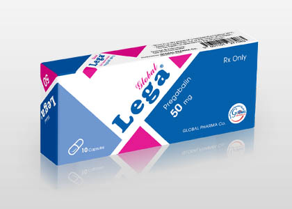
Management with gabapentinoids, tricyclic antidepressants, and antiviral therapy if etiology is viral.
Patient Education
Adhere to medication regimen and protect the affected skin area from mechanical irritation.
Systemic & Specialized Examinations
EN: S1, S2 present. No murmurs. AR: صوتا القلب الأول والثاني طبيعيان. لا توجد نفخات.
EN: Lungs clear to auscultation. AR: الرئتان صافيتان عند التسمع.
EN: Abdomen soft, non-tender. AR: البطن لين ولا يوجد ألم.
EN: Sensory testing reveals allodynia and hyperalgesia in the affected dermatome. AR: يكشف الفحص الحسي عن ألم خيفي وفرط تألم في المنطقة الجلدية المتأثرة.
EN: Unremarkable or not routinely indicated. AR: طبيعي أو غير مطلوب روتينياً.
EN: Unremarkable or not routinely indicated. AR: طبيعي أو غير مطلوب روتينياً.
EN: Unremarkable or not routinely indicated. AR: طبيعي أو غير مطلوب روتينياً.
EN: Unremarkable or not routinely indicated. AR: طبيعي أو غير مطلوب روتينياً.
EN: Unremarkable or not routinely indicated. AR: طبيعي أو غير مطلوب روتينياً.
Comprehensive Clinical Guide: Dorsal Root Ganglionitis (DRGitis)
1. Introduction and Clinical Overview
Dorsal Root Ganglionitis, often clinically referred to as DRGitis, represents a complex inflammatory condition affecting the dorsal root ganglia (DRG)—the sensory relay stations located in the intervertebral foramina of the spinal column. The DRG contains the cell bodies of primary sensory neurons, serving as a critical bottleneck for pain, temperature, and proprioceptive information traveling from the periphery to the central nervous system (CNS).
When these structures become inflamed, the resulting pathology is not merely localized pain, but a profound alteration in neuronal excitability, leading to chronic neuropathic pain syndromes. Unlike standard radiculopathy, which is often mechanical (e.g., disc herniation), DRGitis is primarily neuro-inflammatory or infectious, often resulting in hyperalgesia and allodynia that defy standard conservative treatments.
2. Etiology and Pathophysiology
The etiology of DRGitis is multifactorial, ranging from viral reactivation to autoimmune insults. Understanding the pathophysiology requires a deep dive into the unique environment of the DRG. Unlike the peripheral nerves, the DRG lacks a robust blood-nerve barrier, making it uniquely susceptible to circulating systemic factors, toxins, and pathogens.
Primary Etiological Categories:
- Viral Pathogenesis: The most classic example is Varicella-Zoster Virus (VZV), which remains latent in the DRG following chickenpox and reactivates as Herpes Zoster.
- Autoimmune/Inflammatory: Paraneoplastic syndromes, Sjögren’s syndrome, and systemic lupus erythematosus (SLE) can trigger ganglioneuritis.
- Toxic/Metabolic: Chronic exposure to heavy metals or severe diabetic neuropathy can induce inflammatory changes within the ganglia.
- Mechanical/Post-Surgical: Chronic nerve root compression can induce secondary inflammatory cytokine cascades within the DRG.
Pathophysiological Mechanisms:
- Cytokine Storm: The upregulation of pro-inflammatory cytokines (IL-1β, IL-6, and TNF-α) by satellite glial cells (SGCs) within the DRG.
- Ion Channel Remodeling: Increased expression of voltage-gated sodium channels (specifically Nav1.7 and Nav1.8) on the sensory neuron membranes, lowering the threshold for action potential firing.
- Ectopic Discharge: Spontaneous firing of sensory neurons, which the brain interprets as pain, even in the absence of peripheral stimulus.
- Central Sensitization: Sustained barrage of signals from the DRG leads to long-term potentiation in the spinal cord dorsal horn.
3. Clinical Staging and Grading
Clinical assessment of DRGitis typically follows a functional impairment scale to guide interventional management.
| Stage | Severity | Clinical Manifestations | Functional Impact |
|---|---|---|---|
| Stage I | Mild | Intermittent paresthesia, mild dysesthesia. | Minimal impact on ADLs. |
| Stage II | Moderate | Constant burning, localized allodynia. | Avoidance of certain physical activities. |
| Stage III | Severe | Severe neuropathic pain, motor weakness, sleep disruption. | Significant disability, inability to work. |
| Stage IV | Chronic/Refractory | Constant pain, autonomic dysfunction, muscle atrophy. | Total loss of function, psychological distress. |
4. Standard Presentation and Diagnostic Evaluation
Clinical Presentation
Patients typically present with a "dermatomal" pain distribution. Key symptoms include:
* Allodynia: Pain elicited by non-painful stimuli (e.g., light touch of clothing).
* Hyperalgesia: Exaggerated pain response to painful stimuli.
* Dysesthesia: Unpleasant, abnormal sensations (burning, crawling, electric shocks).
* Loss of Sensory Modality: Often detected in the late stages of inflammatory damage.
Diagnostic Testing Protocol
- MRI with Contrast (High-Resolution): The gold standard for visualizing nerve root inflammation. Contrast enhancement (Gadolinium) of the DRG is highly suggestive of active ganglionitis.
- Electrodiagnostic Studies (EMG/NCS): Used primarily to rule out peripheral neuropathy or radiculopathy. Note: DRGitis may show normal NCS if the lesion is purely sensory.
- Serological Screening: Necessary to rule out underlying autoimmune or infectious causes (e.g., ANA, RF, ESR, CRP, VZV titers).
- Diagnostic DRG Block: A selective anesthetic injection under fluoroscopy. If pain resolves significantly, the DRG is confirmed as the pain generator.
5. Differential Diagnosis
Differentiating DRGitis from other spinal pathologies is critical to avoid unnecessary surgery.
- Lumbar/Cervical Radiculopathy: Usually mechanical. Often improves with decompression; DRGitis does not.
- Post-Herpetic Neuralgia (PHN): A specific, sequela-based form of DRGitis.
- Peripheral Polyneuropathy: Typically distal and symmetric ("stocking-glove"), whereas DRGitis is typically dermatomal.
- Complex Regional Pain Syndrome (CRPS): Often involves DRG involvement but presents with profound autonomic and trophic changes.
6. Risks, Contraindications, and Management
Management must be multimodal. Standard anti-inflammatories (NSAIDs) are often insufficient for the neuropathic component.
Pharmacological Management:
- Gabapentinoids: (Gabapentin/Pregabalin) to modulate ion channel activity.
- Tricyclic Antidepressants: (Amitriptyline/Nortriptyline) to enhance descending inhibitory pathways.
- Topical Agents: Lidocaine 5% patches or Capsaicin 8% for localized relief.
Interventional Contraindications:
- Active Infection: Systemic or local site infection.
- Coagulopathy: Risk of spinal hematoma during diagnostic injections.
- Pregnancy: Relative contraindication for advanced imaging and certain systemic medications.
7. Long-Term Prognosis
The prognosis for DRGitis varies based on the underlying etiology.
* Acute Viral (VZV): Favorable with early antiviral intervention (within 72 hours).
* Autoimmune: Requires long-term disease-modifying antirheumatic drugs (DMARDs).
* Chronic/Idiopathic: Often requires neuromodulation, such as DRG Stimulation (DRG-S), which has shown superior efficacy compared to traditional Spinal Cord Stimulation (SCS) for focal neuropathic pain.
8. Frequently Asked Questions (FAQ)
1. Is Dorsal Root Ganglionitis the same as a pinched nerve?
No. A pinched nerve is usually mechanical compression, while DRGitis is an inflammatory or infectious process affecting the sensory cell bodies.
2. Can DRGitis cause permanent nerve damage?
Yes. If inflammation is left untreated, it can lead to neuronal cell death, resulting in permanent sensory loss or chronic intractable pain.
3. What is the most effective imaging test?
A high-resolution MRI of the spine with Gadolinium contrast is the most effective way to visualize the inflammatory changes within the ganglia.
4. Why don't standard painkillers work for this condition?
Standard NSAIDs target prostaglandins, whereas DRGitis pain is driven by ion channel remodeling and neuro-inflammation that requires neuromodulators like gabapentinoids.
5. Is surgery an option for DRGitis?
Standard decompression surgery is rarely effective. However, neuromodulation (DRG Stimulation) is a highly effective surgical intervention for refractory cases.
6. Can stress trigger a flare-up of DRGitis?
Yes. Stress induces systemic inflammation and sympathetic nervous system activation, which can exacerbate the excitability of inflamed sensory neurons.
7. How long does the diagnostic DRG block last?
A diagnostic block typically provides temporary relief (hours to days). Its primary purpose is to confirm the location of the pain source.
8. Is DRGitis contagious?
Only if the underlying cause is a viral infection, such as the Varicella-Zoster Virus (shingles).
9. Can dietary changes help with inflammation?
While not a primary treatment, an anti-inflammatory diet may help manage systemic cytokine levels, which can indirectly support recovery.
10. What is the role of the satellite glial cells in this condition?
Satellite glial cells surround the neurons in the DRG. When activated by inflammation, they release pro-inflammatory mediators that directly sensitize the neurons, driving the pain cycle.
9. Conclusion
Dorsal Root Ganglionitis is a sophisticated clinical diagnosis that requires a transition from mechanical spinal thinking to neuro-inflammatory management. By targeting the DRG—the true "gateway" of sensory information—clinicians can provide more precise, effective, and long-term relief for patients suffering from this debilitating condition. Early recognition, coupled with appropriate diagnostic imaging and a multimodal treatment approach, remains the gold standard for clinical success.
Disclaimer: This guide is for educational purposes for healthcare professionals and clinical students. It does not replace professional medical judgment. Always refer to current institutional protocols and clinical guidelines when treating patients.