Clinical Assessment & Protocol
Typical Presentation (HPI)
EN: Patient presents with a 15% TBSA full-thickness (third-degree) burn sustained via [thermal/chemical/electrical] mechanism. Patient reports [pain/anesthesia] at the site, with clinical presentation of leathery, charred, or pearly-white eschar. No immediate signs of airway compromise or inhalation injury noted. AR: حضر المريض مصاباً بحروق من الدرجة الثالثة (كاملة السماكة) بنسبة 15% من مساحة سطح الجسم الكلية، ناتجة عن [حراري/كيميائي/كهربائي]. يشكو المريض من [ألم/فقدان حس] في منطقة الإصابة، مع وجود مظهر جلدي متفحم أو أبيض لؤلؤي (eschar). لا توجد علامات سريرية فورية لانسداد مجرى الهواء أو إصابات استنشاقية.
General Examination
EN: Physical examination reveals a 15% TBSA full-thickness burn characterized by non-blanching, insensate, leathery eschar. Surrounding tissue exhibits signs of edema. Capillary refill is absent in the affected area. Peripheral pulses are [palpable/diminished] distal to the injury. AR: يكشف الفحص السريري عن حروق من الدرجة الثالثة بنسبة 15% من مساحة سطح الجسم، تتميز بوجود نسيج متفحم (eschar) غير قابل للتبييض عند الضغط، وفاقد للحس، وذو ملمس جلدي. الأنسجة المحيطة تظهر علامات وذمة. لا يوجد امتلاء شعري في المنطقة المصابة. النبض المحيطي [محسوس/ضعيف] في المناطق البعيدة عن الإصابة.
Treatment Protocol
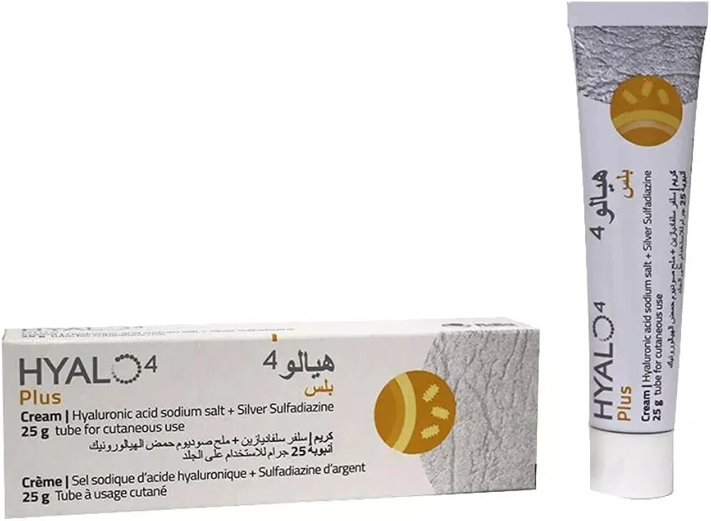
EN: Immediate management includes aggressive fluid resuscitation per Parkland formula, wound debridement, and application of topical antimicrobial agents (e.g., silver sulfadiazine or mafenide acetate). Surgical consultation for early tangential excision and autografting is planned. Pain management and tetanus prophylaxis initiated. AR: تشمل الخطة العلاجية الفورية الإنعاش بالسوائل الوريدية المكثفة وفقاً لمعادلة باركلاند، تنضير الجرح، وتطبيق مضادات ميكروبية موضعية (مثل سلفاديازين الفضة أو أسيتات المافينيد). تم طلب استشارة جراحية لإجراء استئصال مبكر للأنسجة الميتة وزراعة الجلد الذاتي. تم البدء في بروتوكول تسكين الألم والوقاية من التيتانوس.
Patient Education
EN: Patient/family educated on the severity of full-thickness burns, emphasizing the necessity of surgical intervention for healing. Instructions provided on signs of infection (fever, increased erythema, purulent discharge), importance of nutritional support for wound healing, and long-term scar management/physical therapy expectations. AR: تم توعية المريض وذويه بخطورة حروق الدرجة الثالثة، مع التأكيد على ضرورة التدخل الجراحي للالتئام. تم تقديم تعليمات حول علامات العدوى (حمى، زيادة الاحمرار، إفرازات قيحية)، وأهمية الدعم الغذائي لالتئام الجروح، وتوقعات العلاج الطبيعي وإدارة الندبات على المدى الطويل.
Systemic & Specialized Examinations
EN: Unremarkable. Systemic examination is not the primary focus for this reconstructive or aesthetic presentation. AR: طبيعي. الفحص الجهازي ليس التركيز الأساسي لهذه الحالة التجميلية أو الترميمية.
EN: Unremarkable. Systemic examination is not the primary focus for this reconstructive or aesthetic presentation. AR: طبيعي. الفحص الجهازي ليس التركيز الأساسي لهذه الحالة التجميلية أو الترميمية.
EN: Unremarkable. Systemic examination is not the primary focus for this reconstructive or aesthetic presentation. AR: طبيعي. الفحص الجهازي ليس التركيز الأساسي لهذه الحالة التجميلية أو الترميمية.
EN: Unremarkable. Systemic examination is not the primary focus for this reconstructive or aesthetic presentation. AR: طبيعي. الفحص الجهازي ليس التركيز الأساسي لهذه الحالة التجميلية أو الترميمية.
EN: Burn Assessment: ~15% Total Body Surface Area (TBSA) involved. Anterior chest and right arm display leathery, white/charred eschar with absent capillary refill and lack of pinprick sensation. Circumferential burn on the right forearm raises concern for compartment syndrome. AR: تقييم الحرق: حوالي 15% من مساحة سطح الجسم. الصدر الأمامي والذراع الأيمن يظهران قشرة متفحمة/بيضاء قاسية مع غياب التروية الدموية وفقدان الإحساس بالوخز. الحرق المحيطي في الساعد الأيمن يثير القلق من متلازمة الحيز.
EN: Unremarkable. Systemic examination is not the primary focus for this reconstructive or aesthetic presentation. AR: طبيعي. الفحص الجهازي ليس التركيز الأساسي لهذه الحالة التجميلية أو الترميمية.
EN: Unremarkable. Systemic examination is not the primary focus for this reconstructive or aesthetic presentation. AR: طبيعي. الفحص الجهازي ليس التركيز الأساسي لهذه الحالة التجميلية أو الترميمية.
EN: Unremarkable. Systemic examination is not the primary focus for this reconstructive or aesthetic presentation. AR: طبيعي. الفحص الجهازي ليس التركيز الأساسي لهذه الحالة التجميلية أو الترميمية.
EN: Unremarkable. Systemic examination is not the primary focus for this reconstructive or aesthetic presentation. AR: طبيعي. الفحص الجهازي ليس التركيز الأساسي لهذه الحالة التجميلية أو الترميمية.
Orthopedic & Trauma Assessments
EN: Unremarkable. Systemic examination is not the primary focus for this reconstructive or aesthetic presentation. AR: طبيعي. الفحص الجهازي ليس التركيز الأساسي لهذه الحالة التجميلية أو الترميمية.
EN: Unremarkable. Systemic examination is not the primary focus for this reconstructive or aesthetic presentation. AR: طبيعي. الفحص الجهازي ليس التركيز الأساسي لهذه الحالة التجميلية أو الترميمية.
EN: Unremarkable. Systemic examination is not the primary focus for this reconstructive or aesthetic presentation. AR: طبيعي. الفحص الجهازي ليس التركيز الأساسي لهذه الحالة التجميلية أو الترميمية.
EN: Unremarkable. Systemic examination is not the primary focus for this reconstructive or aesthetic presentation. AR: طبيعي. الفحص الجهازي ليس التركيز الأساسي لهذه الحالة التجميلية أو الترميمية.
EN: Unremarkable. Systemic examination is not the primary focus for this reconstructive or aesthetic presentation. AR: طبيعي. الفحص الجهازي ليس التركيز الأساسي لهذه الحالة التجميلية أو الترميمية.
EN: Unremarkable. Systemic examination is not the primary focus for this reconstructive or aesthetic presentation. AR: طبيعي. الفحص الجهازي ليس التركيز الأساسي لهذه الحالة التجميلية أو الترميمية.
EN: Unremarkable. Systemic examination is not the primary focus for this reconstructive or aesthetic presentation. AR: طبيعي. الفحص الجهازي ليس التركيز الأساسي لهذه الحالة التجميلية أو الترميمية.
EN: Unremarkable. Systemic examination is not the primary focus for this reconstructive or aesthetic presentation. AR: طبيعي. الفحص الجهازي ليس التركيز الأساسي لهذه الحالة التجميلية أو الترميمية.
EN: Unremarkable. Systemic examination is not the primary focus for this reconstructive or aesthetic presentation. AR: طبيعي. الفحص الجهازي ليس التركيز الأساسي لهذه الحالة التجميلية أو الترميمية.
Understanding Full-Thickness (Third-Degree) Burns: A Comprehensive Medical Guide
Executive Overview: Definition and Significance
A full-thickness burn, also known as a third-degree burn, represents the most severe form of thermal injury, extending through all layers of the skin – the epidermis and dermis – and potentially into the subcutaneous fat, muscle, or even bone. When such a burn encompasses 15% of the Total Body Surface Area (TBSA), it is classified as a major burn, signifying a critical medical condition requiring immediate and specialized intervention. These injuries are characterized by the complete destruction of dermal elements, including hair follicles, sweat glands, and nerve endings, leading to a lack of pain sensation at the burn site itself. The management of a 15% TBSA third-degree burn is complex, involving a multidisciplinary approach spearheaded by burn surgeons, plastic and reconstructive surgeons, intensivists, and rehabilitation specialists. The primary goals are to achieve wound closure, prevent infection, minimize scarring and contracture, and restore maximal functional and aesthetic outcomes.
Detailed Pathophysiology, Etiology, and Risk Factors
Pathophysiology:
The pathophysiology of a full-thickness burn is characterized by the widespread destruction of cellular structures and the disruption of the skin's barrier function. Thermal energy denatures proteins, leading to cellular death and coagulation necrosis. The depth of the burn is the primary determinant of its severity.
- Epidermal Destruction: The outermost layer of skin is completely ablated.
- Dermal Necrosis: The dermis, containing vital structures like blood vessels, nerves, and appendages, is destroyed. This leads to the absence of pain receptors in the immediate burn area.
- Subcutaneous Tissue Involvement: Depending on the duration and intensity of the heat source, the fat layer beneath the dermis can also be affected. In severe cases, deeper structures like muscle and bone may be exposed.
- Inflammatory Response: A profound systemic inflammatory response is triggered, leading to increased vascular permeability, fluid shifts, and potential organ dysfunction. This can manifest as burn shock, characterized by hypovolemia and reduced cardiac output.
- Infection Risk: The compromised skin barrier is highly susceptible to bacterial colonization and infection, which is a major cause of morbidity and mortality in burn patients. Pathogens like Pseudomonas aeruginosa, Staphylococcus aureus, and Acinetobacter baumannii are common culprits.
- Scarring and Contracture: The body's wound healing process in full-thickness burns relies heavily on fibroblast proliferation and collagen deposition. This often results in hypertrophic scarring and, critically, contractures, which are tightening of the scar tissue that can severely limit range of motion and compromise function, especially around joints.
Etiology:
Full-thickness burns can result from a variety of sources, with the most common being:
- Thermal Burns:
- Flame: Open fires, explosions, flammable liquids.
- Contact: Hot objects (e.g., stoves, irons, radiators), hot liquids (e.g., boiling water, hot oil).
- Scalds: Immersion in or splashing of hot liquids.
- Chemical Burns: Exposure to strong acids (e.g., sulfuric acid, hydrochloric acid) or alkalis (e.g., lye, ammonia). Alkalis are particularly dangerous as they cause deeper, progressive tissue damage.
- Electrical Burns: Passage of electrical current through the body. These burns often have a small entry and exit wound but can cause extensive internal tissue damage, including muscle and nerve injury.
- Radiation Burns: Overexposure to ionizing radiation (e.g., radiotherapy) or UV radiation (severe sunburn).
Risk Factors:
Several factors can increase an individual's risk of sustaining or experiencing more severe outcomes from a full-thickness burn:
- Age: Very young children and the elderly are more vulnerable due to thinner skin and reduced physiological reserves.
- Pre-existing Medical Conditions: Diabetes, peripheral vascular disease, immunocompromise, and respiratory conditions can impair healing and increase complication rates.
- Occupational Hazards: Workers in industries involving high temperatures, flammable materials, or electricity.
- Environmental Factors: Inadequate safety measures in homes or workplaces, living in areas with high incidence of fires or industrial accidents.
- Substance Abuse: Impaired judgment due to alcohol or drug use can lead to accidents.
- Socioeconomic Factors: Poverty can be associated with unsafe living conditions and limited access to preventative measures.
Signs, Symptoms, and Clinical Presentation
The clinical presentation of a full-thickness burn is distinct and often alarming. The absence of pain at the burn site is a key differentiator from superficial burns.
- Appearance: The burn area typically appears dry, leathery, white, charred, or dark brown/black. It may be waxy or opaque.
- Texture: The skin feels inelastic and firm to the touch.
- Sensation: There is a complete absence of pain or sensation in the immediate burn area due to the destruction of nerve endings. However, surrounding areas of partial-thickness burn will be exquisitely painful.
- Blisters: Blisters are generally absent in full-thickness burns because the entire epidermis and dermis have been destroyed. If present, they are usually small and ruptured due to the depth of the injury.
- Capillary Refill: Absence of capillary refill when pressure is applied to the burn area.
- Hair and Gland Destruction: Hair follicles and sweat glands are destroyed, meaning hair will not regrow from the affected area, and the skin will not sweat.
- Associated Injuries: Depending on the cause, patients may present with inhalation injury (cough, hoarseness, sooty sputum, facial burns with soot), chemical burns (tissue discoloration, characteristic odor), or electrical burns (entry/exit wounds, muscle damage, cardiac arrhythmias).
The 15% TBSA designation is a critical component of the initial assessment. This percentage is calculated using standardized charts and formulas, such as the Lund-Browder chart or the Rule of Nines, which assign specific percentages to different body regions. For adults, the Rule of Nines is often used for initial estimation:
| Body Region | Percentage (%) |
|---|---|
| Head and Neck | 9 |
| Each Arm | 9 |
| Chest | 18 |
| Abdomen | 18 |
| Back | 18 |
| Each Leg | 18 |
| Perineum | 1 |
For a 15% TBSA burn, this could involve a significant portion of a limb, the entire trunk, or a combination of smaller areas. The extent of the burn directly correlates with the severity of the systemic response and the complexity of management.
Standard Diagnostic Evaluation & Workup
The diagnosis of a full-thickness burn is primarily a clinical one, based on the characteristic appearance and depth. However, a comprehensive diagnostic workup is essential to assess the extent of injury, identify associated complications, and guide treatment.
1. Clinical Assessment:
The initial assessment by experienced medical professionals is paramount. This includes:
- History Taking: Mechanism of injury, duration of exposure, presence of inhalation injury, past medical history, allergies, and medications.
- Physical Examination:
- Burn Depth Classification: Differentiating between superficial, partial-thickness, and full-thickness burns.
- TBSA Estimation: Using the Rule of Nines or Lund-Browder chart.
- Assessment for Inhalation Injury: Examining the face, mouth, and upper airways for signs of soot, burns, or edema.
- Circulatory Assessment: Checking for peripheral pulses and signs of compartment syndrome, especially in circumferential burns.
- Neurological Status: Assessing for any neurological deficits, particularly with electrical burns.
2. Laboratory Assays:
Blood tests are crucial for assessing systemic response and identifying complications.
- Complete Blood Count (CBC): To assess for anemia (due to fluid shifts and blood loss) and leukocytosis (indicating infection or inflammation).
- Electrolytes (Sodium, Potassium, Chloride, Bicarbonate): To monitor for electrolyte imbalances caused by fluid shifts and resuscitation.
- Renal Function Tests (BUN, Creatinine): To assess kidney function, which can be affected by hypovolemia and systemic inflammation.
- Liver Function Tests (LFTs): To monitor for potential hepatic dysfunction.
- Coagulation Studies (PT, PTT, INR): To assess for coagulopathy, which can occur in severe burns.
- Carboxyhemoglobin Levels: If inhalation injury from smoke is suspected.
- Blood Glucose: To monitor for hyperglycemia, a common stress response.
- Blood Cultures: If infection is suspected.
3. Imaging Studies:
Imaging plays a role in diagnosing associated injuries and complications.
- Chest X-ray: To evaluate for pulmonary edema, pneumonia, or other signs of smoke inhalation injury.
- Computed Tomography (CT) Scan:
- CT Angiography: To assess for vascular compromise or injury, especially in deep burns or those with suspected arterial involvement.
- CT of the Airway: If significant inhalation injury is suspected, to assess for tracheal or bronchial edema.
- CT of the Spine/Abdomen/Pelvis: If trauma is suspected as a co-factor in the burn injury (e.g., falls).
- Electrocardiogram (ECG): Essential for all patients with significant burns, especially electrical burns, to detect arrhythmias or cardiac injury.
4. Biopsy (Less Common for Initial Diagnosis):
While not typically performed for initial diagnosis of a full-thickness burn, a biopsy could be considered in ambiguous cases where the depth of the burn is uncertain and the information would significantly alter management. Histopathology would reveal complete necrosis of all dermal elements. However, the characteristic clinical appearance is usually sufficient for diagnosis.
5. Wound Cultures:
Routine wound cultures are generally not recommended in the initial phase unless there are signs of clinical infection. However, if infection is suspected, a wound swab or biopsy for culture and sensitivity testing is performed to identify the causative organism and guide antibiotic therapy.
Therapeutic Interventions
The management of a 15% TBSA full-thickness burn is a critical, time-sensitive endeavor requiring a multidisciplinary approach. Treatment focuses on immediate resuscitation, wound management, infection control, and long-term rehabilitation.
1. Resuscitation:
This is the cornerstone of early management for major burns.
- Fluid Resuscitation: Intravenous fluid administration is crucial to combat burn shock and maintain adequate organ perfusion. The Parkland formula is a common guideline for calculating initial fluid needs (e.g., 4 mL of Lactated Ringer's solution per kilogram of body weight per percentage of TBSA burned). This fluid is typically administered over the first 24 hours, with half given in the first 8 hours and the remainder over the next 16 hours. Close monitoring of urine output and hemodynamic parameters guides ongoing fluid management.
- Airway Management: For patients with suspected inhalation injury, airway protection (e.g., intubation) may be necessary.
2. Pharmacotherapy:
- Analgesia: Despite the absence of pain in the full-thickness burn itself, surrounding partial-thickness burns and procedures (e.g., wound care, debridement) are painful. Opioids (e.g., morphine, fentanyl) are typically used for pain management.
- Antibiotics: Prophylactic systemic antibiotics are generally not recommended. Antibiotics are reserved for established infections, guided by wound cultures and sensitivity testing. Topical antimicrobial agents (e.g., silver sulfadiazine, mafenide acetate, bacitracin) are used to prevent bacterial colonization of the burn wound.
- Tetanus Prophylaxis: Tetanus toxoid should be administered if the patient's immunization status is not up-to-date.
3. Surgical Interventions:
- Early Debridement: The removal of necrotic tissue (eschar) is essential to prevent bacterial proliferation and prepare the wound bed for grafting. This can be done surgically (excision) or enzymatically.
- Wound Excision and Grafting: This is the definitive treatment for full-thickness burns.
- Tangential Excision: The burned tissue is surgically shaved off layer by layer until healthy, bleeding tissue is reached. This is typically performed within the first few days after injury.
- Skin Grafting: Once the wound bed is prepared, it is covered with a skin graft.
- Autograft: Skin taken from an unburned area of the patient's own body. This is the gold standard for definitive wound closure. Split-thickness skin grafts (STSG) are most commonly used, where a thin layer of epidermis and a portion of the dermis are harvested. Full-thickness skin grafts may be used for smaller areas or cosmetically sensitive regions.
- Allograft: Temporary coverage using skin from a deceased donor.
- Xenograft: Temporary coverage using skin from an animal (e.g., pig).
- Skin Substitutes: Cultured epithelial autografts or various bioengineered skin substitutes may be used in extensive burns or when autograft donor sites are limited.
- Escharotomy and Fasciotomy: For circumferential full-thickness burns (encircling a limb or the chest), escharotomy (incisions through the eschar) or fasciotomy (incision through the fascia) may be required to relieve pressure, restore circulation, and allow for adequate chest wall excursion for breathing.
4. Wound Care and Dressings:
Regular wound cleaning, debridement of loose tissue, and application of appropriate dressings are critical. Dressings help to maintain a moist wound environment, protect the graft, absorb exudate, and prevent infection.
5. Nutritional Support:
Burn patients have significantly increased metabolic demands. Adequate protein and calorie intake is vital for wound healing and immune function. Enteral or parenteral nutrition may be required.
6. Lifestyle and Rehabilitation:
- Physical Therapy: Early mobilization and range-of-motion exercises are crucial to prevent contractures and maintain function.
- Occupational Therapy: To assist with activities of daily living and regaining independence.
- Psychological Support: Burn injuries have a profound psychological impact. Counseling and support groups are essential for patients and their families to cope with pain, disfigurement, and the long recovery process.
- Scar Management: Once grafts have healed, scar management techniques such as silicone sheeting, pressure garments, massage, and laser therapy are used to improve scar pliability and reduce hypertrophy.
Frequently Asked Questions (FAQ)
1. What is the most common cause of third-degree burns?
The most common causes of third-degree burns are thermal injuries, including prolonged contact with flames, hot objects, or hot liquids (scalds). Chemical and electrical burns also contribute significantly.
2. Does a third-degree burn hurt?
The full-thickness burn itself, where all nerve endings are destroyed, is typically painless. However, the surrounding areas of partial-thickness burn will be extremely painful, and procedures like wound cleaning and dressing changes can also cause significant pain.
3. How long does it take to recover from a 15% TBSA third-degree burn?
Recovery is a lengthy and complex process. Initial hospitalization can last for weeks to months, depending on the need for surgery and the presence of complications. Full functional and cosmetic recovery can take years, involving multiple reconstructive surgeries, extensive physical therapy, and ongoing scar management.
4. What is the main risk associated with a third-degree burn?
The primary risks associated with third-degree burns are infection, severe scarring leading to contractures and loss of function, and significant fluid and electrolyte imbalances during the initial resuscitation phase.
5. Will I have permanent scarring after a third-degree burn?
Yes, third-degree burns will always result in permanent scarring. The goal of treatment is to minimize the severity of the scarring and its impact on function through grafting and subsequent scar management techniques.
6. What are the signs that a burn is a third-degree burn?
Key signs include a dry, leathery, white, charred, or dark brown appearance; absence of pain in the burn area; and destruction of all skin layers, meaning no hair follicles or sweat glands are present.
7. What is TBSA and why is 15% significant?
TBSA stands for Total Body Surface Area. A 15% TBSA burn is considered a major burn, indicating a significant injury that triggers a systemic inflammatory response and requires specialized, intensive medical care, often in a dedicated burn center.
8. What is the standard treatment for a third-degree burn?
The standard treatment involves immediate fluid resuscitation, surgical debridement of the burned tissue, and coverage of the wound with skin grafts (autografting). Pain management, infection control, and nutritional support are also critical components.
9. Can a third-degree burn heal on its own?
No, a third-degree burn cannot heal on its own. Because all layers of the skin, including the cells that regenerate skin, are destroyed, the wound requires surgical intervention to close the defect, typically with skin grafts.
10. What are the long-term complications of a third-degree burn?
Long-term complications can include severe disfigurement, chronic pain, loss of sensation, contractures limiting movement, itching (pruritus), increased sensitivity to temperature, and an increased risk of skin cancer developing in the scar tissue over many years.