Clinical Assessment & Protocol
Typical Presentation (HPI)
EN: Patient presents with chronic, progressive right shoulder pain, localized to the glenohumeral joint. Symptoms are exacerbated by overhead activity and internal rotation, with associated stiffness and mechanical symptoms including crepitus. Pain is worse with activity and improves slightly with rest. No history of acute trauma, instability, or neurological deficits. AR: يعاني المريض من ألم مزمن ومتفاقم في الكتف الأيمن، يتركز في المفصل الحقاني العضدي. تزداد الأعراض سوءاً مع الأنشطة التي تتطلب رفع الذراع فوق مستوى الرأس والحركات الدورانية الداخلية، مع وجود تيبس وأعراض ميكانيكية تشمل فرقعة المفصل. يزداد الألم مع النشاط ويتحسن قليلاً مع الراحة. لا يوجد تاريخ لصدمة حادة، أو عدم استقرار في المفصل، أو عجز عصبي.
General Examination
EN: Right shoulder examination reveals atrophy of the supraspinatus and infraspinatus fossae. Tenderness to palpation noted over the glenohumeral joint line. Active and passive range of motion is significantly restricted in all planes, particularly abduction and external rotation. Crepitus is palpable during passive motion. Neurovascular status is intact distally. AR: يكشف فحص الكتف الأيمن عن ضمور في الحفرة فوق الشوكة وتحت الشوكة. لوحظ وجود ألم عند الجس على طول خط المفصل الحقاني العضدي. مدى الحركة النشط والسلبي محدود بشكل كبير في جميع المستويات، وخاصة في حركات الإبعاد والدوران الخارجي. لوحظ وجود فرقعة ملموسة أثناء الحركة السلبية. الحالة العصبية الوعائية سليمة في الأطراف البعيدة.
Treatment Protocol
EN: Initiate conservative management including activity modification, physical therapy focusing on range of motion and periscapular strengthening, and non-steroidal anti-inflammatory drugs (NSAIDs). Consider intra-articular corticosteroid or hyaluronic acid injection for symptomatic relief. Discuss surgical options, including arthroscopic debridement or total shoulder arthroplasty, if conservative measures fail. AR: البدء بالعلاج التحفظي الذي يشمل تعديل الأنشطة، والعلاج الطبيعي الذي يركز على مدى الحركة وتقوية العضلات حول لوح الكتف، واستخدام مضادات الالتهاب غير الستيرويدية. النظر في حقن الكورتيكوستيرويد أو حمض الهيالورونيك داخل المفصل لتخفيف الأعراض. مناقشة الخيارات الجراحية، بما في ذلك التنظير المفصلي أو استبدال مفصل الكتف بالكامل، في حال فشل الإجراءات التحفظية.
Patient Education
EN: Glenohumeral osteoarthritis is a degenerative condition involving the wearing down of the articular cartilage in the shoulder joint. Focus on maintaining joint mobility through prescribed exercises and avoiding high-impact activities that aggravate pain. Monitor for worsening weakness or night pain, and adhere to the physical therapy regimen to optimize functional outcomes. AR: خشونة المفصل الحقاني العضدي هي حالة تنكسية تتضمن تآكل الغضروف المفصلي في مفصل الكتف. يجب التركيز على الحفاظ على مرونة المفصل من خلال التمارين الموصوفة وتجنب الأنشطة ذات التأثير العالي التي تزيد من حدة الألم. يرجى مراقبة أي تفاقم في الضعف أو ألم ليلي، والالتزام ببرنامج العلاج الطبيعي لتحسين النتائج الوظيفية.
Systemic & Specialized Examinations
EN: Distal neurovascular status intact globally. AR: الحالة العصبية والوعائية الطرفية سليمة تماماً.
Orthopedic & Trauma Assessments
EN: Insidious degenerative wear and tear. No acute trauma. AR: تآكل تنكسي تدريجي. لا توجد صدمة حادة.
EN: Antalgic gait. Reduced stance phase on the affected side. Trendelenburg or varus thrust may be present. AR: مشية متألمة. قصر في مرحلة الوقوف على الجانب المصاب. قد يوجد اندفاع تقوسي أو علامة ترندلينبورغ.
EN: Moderate chronic joint effusion/thickening. Obvious malalignment in the coronal plane. Mild surrounding muscle atrophy. AR: انصباب/تسمك مفصلي مزمن. سوء محاذاة واضح. ضمور خفيف في العضلات المحيطة.
EN: Grind tests (Patellar/FABER) strongly positive. Ligament tests negative. AR: اختبارات الطحن (مثل FABER) إيجابية بقوة. اختبارات الأربطة سلبية.
EN: 4/5 strength in proximal muscles due to pain inhibition. Distal strength 5/5. AR: قوة 4/5 في العضلات القريبة بسبب تثبيط الألم. القوة الطرفية 5/5.
EN: Sensation intact to light touch in all dermatomes. AR: الإحساس سليم للمس الخفيف في جميع التوزيعات العصبية.
EN: 2+ symmetric deep tendon reflexes. AR: المنعكسات العميقة 2+ ومتماثلة.
EN: DP and PT pulses 2+ bounding. Capillary refill < 2 seconds. AR: نبضات القدم 2+ قوية. عودة امتلاء الشعيرات < ثانيتين.
Comprehensive Clinical Guide: Glenohumeral Osteoarthritis (Right Shoulder)
1. Introduction and Clinical Overview
Glenohumeral osteoarthritis (GHOA) represents a degenerative condition characterized by the progressive erosion of the articular cartilage within the glenohumeral joint. While the hip and knee are more frequently associated with weight-bearing osteoarthritis, the glenohumeral joint is uniquely susceptible to degenerative changes due to its high mobility and the complex biomechanical interplay between the humeral head and the glenoid fossa.
When localized to the right shoulder, the condition frequently impacts the dominant extremity, leading to significant functional impairment in activities of daily living (ADLs), occupational performance, and athletic pursuits. As the cartilage wears thin, the subchondral bone is exposed, leading to osteophyte formation, joint space narrowing, and chronic synovial inflammation.
2. Etiology and Pathophysiology
The degradation of the glenohumeral joint is rarely a result of a single event; rather, it is a multi-factorial process involving mechanical, biochemical, and genetic components.
Primary Etiology
- Primary Osteoarthritis: Idiopathic, often associated with age-related wear and tear.
- Post-Traumatic: Sequelae of previous fractures (proximal humerus), dislocations, or chronic instability.
- Inflammatory Arthropathies: Rheumatoid arthritis or ankylosing spondylitis.
- Crystalline Arthropathies: Gout or pseudogout (CPPD).
- Rotator Cuff Tear Arthropathy (RCTA): Chronic, massive rotator cuff tears leading to proximal migration of the humeral head and subsequent cartilage destruction.
- Avascular Necrosis (AVN): Compromised blood supply to the humeral head.
Pathophysiological Mechanisms
The breakdown of the extracellular matrix (ECM) of the articular cartilage is driven by an imbalance between anabolic and catabolic processes. Chondrocytes, which typically maintain the matrix, shift toward a catabolic state, secreting matrix metalloproteinases (MMPs) and inflammatory cytokines (IL-1, TNF-alpha). This leads to:
1. Fibrillation: Micro-cracks develop in the superficial layer of the cartilage.
2. Erosion: Progression to full-thickness cartilage loss.
3. Subchondral Sclerosis: Increased bone density resulting from abnormal loading.
4. Osteophytosis: Marginal bone growth as the body attempts to increase surface area to distribute load.
3. Clinical Staging and Grading (Samilson-Prieto Classification)
Clinicians utilize the Samilson-Prieto classification system to quantify the severity of GHOA based on radiographic findings:
| Grade | Clinical Description | Radiographic Features |
|---|---|---|
| Mild | Early stage, minimal symptoms | Osteophytes < 3 mm |
| Moderate | Functional limitation | Osteophytes 3–7 mm; joint space narrowing |
| Severe | Significant pain/stiffness | Osteophytes > 7 mm; severe joint space narrowing |
4. Standard Clinical Presentation
Patients presenting with right-sided GHOA typically report a insidious onset of symptoms.
- Pain: Deep, aching pain localized to the posterior or lateral aspect of the shoulder, often radiating toward the deltoid insertion.
- Stiffness: Progressive loss of both active and passive range of motion (ROM), particularly in external rotation and abduction.
- Crepitus: Audible or palpable grinding during glenohumeral motion.
- Functional Limitations: Difficulty reaching overhead, inability to reach behind the back (internal rotation deficit), and pain when sleeping on the affected side.
- Muscle Atrophy: In chronic cases, disuse atrophy of the supraspinatus and infraspinatus muscles is common.
5. Diagnostic Methodology
A definitive diagnosis of GHOA requires a synthesis of history, physical examination, and imaging.
Key Diagnostic Tests
- Physical Exam:
- Passive ROM assessment: Detection of capsular patterns (typically ER > Abduction > IR).
- Cross-body adduction test: Often positive, suggesting AC joint involvement or GHOA.
- Neer/Hawkins-Kennedy: To rule out subacromial impingement.
- Imaging:
- True AP (Grashey view): Essential to visualize the glenohumeral joint space.
- Axillary Lateral: To assess the relationship between the humeral head and the glenoid.
- MRI (Optional): Utilized primarily to evaluate the status of the rotator cuff and labrum if surgical intervention is planned.
- CT Scan: Often necessary for preoperative planning to assess glenoid bone stock and version.
Differential Diagnosis
- Adhesive Capsulitis (Frozen Shoulder)
- Rotator Cuff Tendinopathy
- Acromioclavicular (AC) Joint Arthritis
- Cervical Radiculopathy (C5-C6)
- Superior Labrum Anterior-to-Posterior (SLAP) Lesions
6. Management and Long-Term Prognosis
Conservative Management (First-Line)
- Physical Therapy: Focus on scapular stabilization, rotator cuff strengthening, and maintenance of ROM.
- Pharmacology: NSAIDs (as tolerated) for inflammation, topical analgesics, and occasional corticosteroid injections.
- Lifestyle Modification: Activity modification to avoid overhead repetitive stress.
Surgical Management
When conservative measures fail to provide adequate relief, surgical intervention is considered:
1. Arthroscopic Debridement: Indicated for early-stage disease with mechanical symptoms.
2. Anatomic Total Shoulder Arthroplasty (TSA): The gold standard for primary GHOA with an intact rotator cuff.
3. Reverse Total Shoulder Arthroplasty (rTSA): Indicated for GHOA associated with massive, irreparable rotator cuff tears (RCTA).
4. Hemiarthroplasty: Reserved for younger patients or specific anatomical conditions where the glenoid cannot be resurfaced.
Long-Term Prognosis
GHOA is a progressive condition. While non-operative management can successfully manage symptoms for years, many patients will eventually require surgical intervention. Post-operative outcomes for TSA are generally excellent, with high patient satisfaction scores and significant restoration of function, provided the rotator cuff remains functional.
7. Risks, Side Effects, and Contraindications
- NSAID Risks: Gastrointestinal bleeding, renal impairment, and cardiovascular events.
- Injection Risks: Infection (septic arthritis is a surgical emergency), post-injection flare, and potential weakening of tendons if over-utilized.
- Surgical Risks: Infection (periprosthetic), neurovascular injury, implant loosening, instability, and stiffness.
- Contraindications for TSA: Active infection, severe deltoid paralysis, or severe bone loss (unless bone grafting is viable).
8. Massive FAQ Section
Q1: Is GHOA the same as "Frozen Shoulder"?
A: No. GHOA is a structural, degenerative condition of the cartilage. Frozen shoulder (adhesive capsulitis) is characterized by thickening and inflammation of the joint capsule. While both cause stiffness, they have different underlying pathologies.
Q2: Will my right shoulder pain eventually require surgery?
A: Not necessarily. Many patients manage symptoms successfully through physical therapy and activity modification. Surgery is usually reserved for cases that significantly limit quality of life.
Q3: Can I keep exercising if I have GHOA?
A: Yes, but modification is key. Low-impact activities like swimming or stationary cycling are encouraged. Avoid heavy overhead pressing or high-impact contact sports that exacerbate joint loading.
Q4: How effective are corticosteroid injections?
A: They are excellent for short-term pain relief and inflammation reduction. However, they do not cure the underlying degeneration and should be used sparingly to avoid potential damage to the surrounding tendons.
Q5: What is the difference between TSA and rTSA?
A: Anatomic TSA replaces the joint surfaces with a ball-and-socket mimicking the natural anatomy. rTSA reverses the anatomy (the ball becomes the glenoid side), which allows the deltoid to compensate for a deficient rotator cuff.
Q6: Does diet affect GHOA?
A: While no specific diet cures GHOA, an anti-inflammatory diet (rich in Omega-3s and antioxidants) and maintaining a healthy weight can reduce systemic inflammation and joint load.
Q7: Can supplements like Glucosamine help?
A: Clinical evidence is mixed. While some patients report subjective improvement, high-quality studies have not definitively proven that these supplements regenerate cartilage.
Q8: How long is the recovery after a total shoulder replacement?
A: Full recovery typically takes 6–12 months. This involves a strictly supervised physical therapy program to protect the subscapularis repair and regain mobility.
Q9: Why is my right shoulder worse than my left?
A: Because the right side is typically the dominant extremity, it is subjected to more frequent use, higher impact loads, and a higher cumulative frequency of micro-trauma throughout a lifetime.
Q10: Can I prevent GHOA?
A: You cannot fully prevent age-related degeneration, but you can minimize risk by avoiding traumatic injuries, maintaining rotator cuff strength, and maintaining good posture to ensure proper glenohumeral kinematics.
9. Conclusion
Glenohumeral osteoarthritis of the right shoulder is a complex, debilitating condition that requires a highly tailored clinical approach. By understanding the biomechanical foundations and the progressive nature of the pathophysiology, clinicians can provide patients with a roadmap from initial diagnosis through long-term management. Early intervention through physical therapy and lifestyle adjustment remains the cornerstone of care, with advanced surgical options available to restore function when conservative limits are reached.
Disclaimer: This document is for informational and educational purposes only and does not constitute medical advice, diagnosis, or treatment. Always seek the advice of a qualified orthopedic surgeon or healthcare provider with any questions regarding a medical condition.
Related Clinical Integration
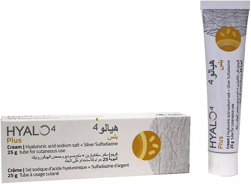
In a modern clinical setting, the management of Glenohumeral Osteoarthritis, Right Shoulder, requires a comprehensive, multidisciplinary approach that integrates evidence-based pharmacotherapy, specialized surgical interventions, and patient-centered education. Initial conservative management often utilizes medications such as Hyalo4 plus cream / هيالو 4 بلس كريم 0.2% / 1% and Celcox / سيلكوكس 100mg alongside the use of a Simple Shoulder Sling (UltraSling) / حمالة كتف بسيطة (ألتراسلينغ) (الأطراف الصناعية والجبائر التقويمية) to manage inflammation and support joint stability. When surgical intervention becomes necessary, our facility employs advanced techniques such as Cemented Hemiarthroplasty for Pathologic Fracture / رأب نصف المفصل الملحوم لكسر مرضي (عملية كبرى في غرف العمليات) or Reverse Total Shoulder Arthroplasty (rTSA) / رأب مفصل الكتف الكلي المعكوس (rTSA) (عملية كبرى في غرف العمليات), supported by precision tools including Trephine Reamer Sets / مجموعات موسعات الثقب (تريفين), Distal Femoral Replacement (Tumor Prosthesis) / استبدال الفخذ البعيد (طرف صناعي للأورام), and the [Oscillating Bone Saw Blade (Wide, Narrow, Deep Cut) / شفرة منشار عظمي متذبذب (عريض، ضيق، قطع عميق)](https://yemenhealthos.com/ar/clinic/instruments/oscillating-bone-saw-blade-wide-