Clinical Assessment & Protocol
Typical Presentation (HPI)
EN: Patient presents with symptoms suggestive of chronic prostatitis, including irritative and obstructive voiding symptoms (dysuria, frequency, urgency, nocturia), perineal or suprapubic discomfort, and occasional hematospermia. History often reveals recurrent urinary tract infections or recent intravesical therapy (e.g., BCG). AR: يعاني المريض من أعراض توحي بالتهاب البروستاتا المزمن، بما في ذلك أعراض بولية تهيجية وانسدادية (عسر تبول، تكرار، إلحاح، تبول ليلي)، مع انزعاج في منطقة العجان أو فوق العانة، ووجود دم في السائل المنوي أحياناً. التاريخ المرضي غالباً ما يكشف عن التهابات متكررة في المسالك البولية أو علاج داخل المثانة حديثاً (مثل لقاح BCG).
General Examination
EN: Digital Rectal Examination (DRE) reveals a firm, indurated, or nodular prostate gland, often mimicking prostatic carcinoma. Tenderness may be present. Absence of fluctuance helps rule out prostatic abscess. Systemic examination is typically unremarkable unless associated with systemic granulomatous disease. AR: فحص البروستاتا عبر المستقيم (DRE) يكشف عن غدة بروستاتية صلبة، متصلبة، أو عقدية، مما يحاكي في كثير من الأحيان سرطان البروستاتا. قد يوجد إيلام عند اللمس. غياب التموج (Fluctuance) يساعد في استبعاد وجود خراج بروستاتي. الفحص السريري العام عادة ما يكون طبيعياً ما لم يكن مرتبطاً بمرض حبيبي جهازي.
Treatment Protocol
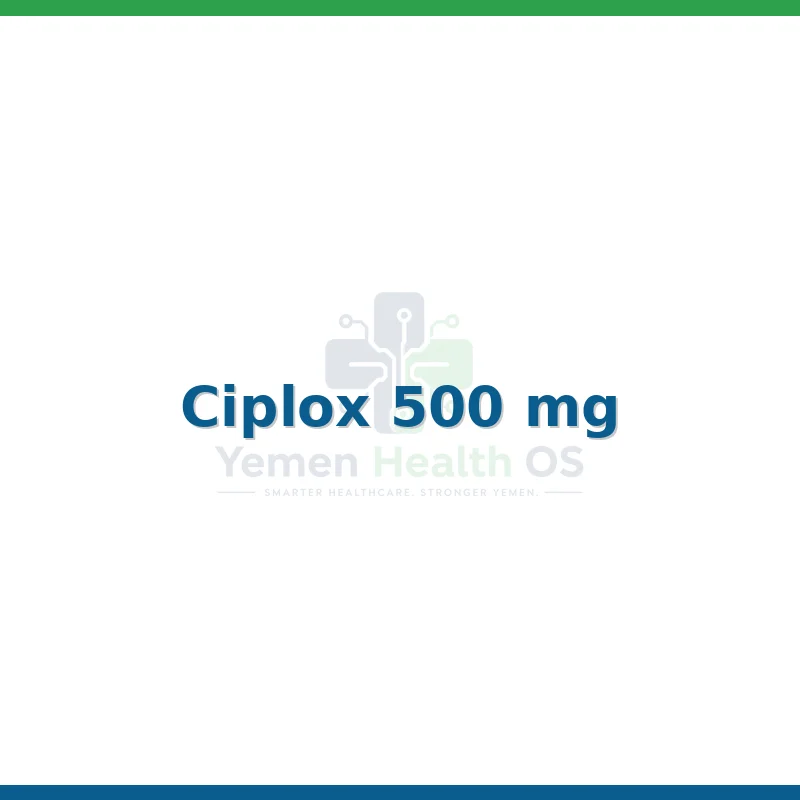
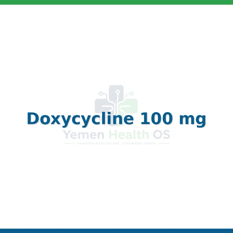
EN: Management involves a multimodal approach: 1) Antibiotic therapy if infection is suspected (e.g., fluoroquinolones for 4-6 weeks). 2) Anti-inflammatory agents (NSAIDs) for pain management. 3) Alpha-blockers to alleviate obstructive voiding symptoms. 4) Corticosteroids may be indicated in severe idiopathic cases. Surgical intervention (TURP) is reserved for refractory cases with significant bladder outlet obstruction. AR: يتضمن العلاج نهجاً متعدد الوسائط: 1) العلاج بالمضادات الحيوية في حال الاشتباه بوجود عدوى (مثل الفلوروكينولونات لمدة 4-6 أسابيع). 2) مضادات الالتهاب غير الستيرويدية للتحكم في الألم. 3) حاصرات ألفا لتخفيف أعراض انسداد المسالك البولية. 4) قد يوصى بالكورتيكوستيرويدات في الحالات مجهولة السبب الشديدة. التدخل الجراحي (استئصال البروستاتا عبر الإحليل) مخصص للحالات المستعصية التي تعاني من انسداد كبير في مخرج المثانة.
Patient Education
EN: Granulomatous prostatitis is a rare inflammatory condition of the prostate that can mimic cancer. It is not a malignancy. You may require periodic follow-up, including PSA monitoring and potentially repeat biopsies, to ensure the condition is resolving and to rule out underlying prostate cancer. Report any worsening of urinary retention or fever immediately. AR: التهاب البروستاتا الحبيبي هو حالة التهابية نادرة في البروستاتا قد تحاكي السرطان في أعراضها، لكنها ليست ورماً خبيثاً. قد تحتاج إلى متابعة دورية، بما في ذلك مراقبة مستضد البروستاتا النوعي (PSA) وربما إعادة أخذ خزعات، للتأكد من تحسن الحالة واستبعاد وجود سرطان بروستاتا كامن. يرجى إبلاغ الطبيب فوراً في حال حدوث أي تدهور في احتباس البول أو ظهور حمى.
Systemic & Specialized Examinations
EN: S1, S2 present. No murmurs. Normal rate and rhythm. AR: صوتا القلب الأول والثاني طبيعيان. لا توجد نفخات.
EN: Lungs clear to auscultation bilaterally. No wheezes or crackles. AR: الرئتان صافيتان عند التسمع. لا يوجد أزيز أو كراكر.
EN: Abdomen and flank examined to rule out upper tract involvement or palpable masses. AR: تم فحص البطن والخاصرة لاستبعاد إصابة الجهاز البولي العلوي أو الكتل الملموسة.
EN: Alert, oriented x3. Normal sacral reflexes (bulbocavernosus intact). AR: واعي ومدرك. المنعكسات العجزية طبيعية.
EN: Unremarkable or not routinely indicated for this specific urological/andrological pathology. AR: طبيعي أو غير مطلوب روتينياً لهذا المرض البولي أو الذكوري.
EN: Unremarkable or not routinely indicated for this specific urological/andrological pathology. AR: طبيعي أو غير مطلوب روتينياً لهذا المرض البولي أو الذكوري.
EN: Unremarkable or not routinely indicated for this specific urological/andrological pathology. AR: طبيعي أو غير مطلوب روتينياً لهذا المرض البولي أو الذكوري.
EN: Unremarkable or not routinely indicated for this specific urological/andrological pathology. AR: طبيعي أو غير مطلوب روتينياً لهذا المرض البولي أو الذكوري.
EN: Unremarkable or not routinely indicated for this specific urological/andrological pathology. AR: طبيعي أو غير مطلوب روتينياً لهذا المرض البولي أو الذكوري.
Orthopedic & Trauma Assessments
EN: Unremarkable or not routinely indicated for this specific urological/andrological pathology. AR: طبيعي أو غير مطلوب روتينياً لهذا المرض البولي أو الذكوري.
EN: Unremarkable or not routinely indicated for this specific urological/andrological pathology. AR: طبيعي أو غير مطلوب روتينياً لهذا المرض البولي أو الذكوري.
EN: Unremarkable or not routinely indicated for this specific urological/andrological pathology. AR: طبيعي أو غير مطلوب روتينياً لهذا المرض البولي أو الذكوري.
EN: Unremarkable or not routinely indicated for this specific urological/andrological pathology. AR: طبيعي أو غير مطلوب روتينياً لهذا المرض البولي أو الذكوري.
EN: Unremarkable or not routinely indicated for this specific urological/andrological pathology. AR: طبيعي أو غير مطلوب روتينياً لهذا المرض البولي أو الذكوري.
EN: Unremarkable or not routinely indicated for this specific urological/andrological pathology. AR: طبيعي أو غير مطلوب روتينياً لهذا المرض البولي أو الذكوري.
EN: Unremarkable or not routinely indicated for this specific urological/andrological pathology. AR: طبيعي أو غير مطلوب روتينياً لهذا المرض البولي أو الذكوري.
EN: Unremarkable or not routinely indicated for this specific urological/andrological pathology. AR: طبيعي أو غير مطلوب روتينياً لهذا المرض البولي أو الذكوري.
EN: Unremarkable or not routinely indicated for this specific urological/andrological pathology. AR: طبيعي أو غير مطلوب روتينياً لهذا المرض البولي أو الذكوري.
Understanding Granulomatous Prostatitis: A Clinical Overview
Granulomatous prostatitis (GP) is a rare, distinct inflammatory condition of the prostate gland characterized by the presence of granulomas—aggregates of activated macrophages, epithelioid cells, and giant cells—within the prostatic parenchyma. Classified under ICD-10 code N41.1_2, it represents a diagnostic challenge for urologists because it frequently mimics prostate carcinoma on digital rectal examination (DRE) and imaging studies.
Unlike common bacterial prostatitis, which is usually infectious, granulomatous prostatitis is often categorized into two main subtypes: Non-specific (idiopathic) and Specific (associated with systemic diseases or infections). Distinguishing between these is the paramount goal of the urological workup to ensure appropriate management and to rule out malignancy.
Pathophysiology, Etiology, and Risk Factors
The formation of a granuloma in the prostate is typically a response to a persistent irritant or an immune-mediated process. The etiology is multifaceted and can be broadly categorized as follows:
Etiological Classifications
- Idiopathic (Non-specific): The most common form, often linked to the extravasation of prostatic secretions into the surrounding stroma, triggering a foreign-body type inflammatory response.
- Infectious: Secondary to systemic infections such as Tuberculosis (Mycobacterium tuberculosis), Fungal infections (e.g., Histoplasmosis, Cryptococcosis), or Brucellosis.
- Post-procedural/Iatrogenic: A common cause following Transurethral Resection of the Prostate (TURP), needle biopsies, or intravesical BCG therapy for bladder cancer.
- Systemic/Autoimmune: Rare associations with conditions like Sarcoidosis, Wegener’s granulomatosis (Granulomatosis with polyangiitis), or Eosinophilic granulomatosis with polyangiitis.
Risk Factors
- Recent Urological Instrumentation: Recent biopsies or surgeries are the most significant risk factors for iatrogenic GP.
- Intravesical BCG Therapy: Patients treated for superficial bladder cancer are at high risk due to the immune stimulation caused by the BCG vaccine.
- Immunocompromised Status: Patients on immunosuppressive therapy or those with HIV/AIDS are more susceptible to infectious granulomatous prostatitis.
- Autoimmune Predisposition: Individuals with a history of systemic vasculitis or sarcoidosis.
| Type | Primary Trigger | Clinical Context |
|---|---|---|
| Non-specific | Extravasation of prostatic fluid | Spontaneous, unknown origin |
| Iatrogenic | BCG therapy / Biopsy | Post-procedural follow-up |
| Infectious | Pathogens (TB, Fungal) | Systemic symptoms, travel history |
| Systemic | Autoimmune vasculitis | Multiorgan involvement |
Signs, Symptoms, and Clinical Presentation
The clinical presentation of granulomatous prostatitis is often indistinguishable from prostate cancer or acute bacterial prostatitis, which is why it is often referred to as a "masquerader."
Common Symptoms
- Lower Urinary Tract Symptoms (LUTS): Frequency, urgency, nocturia, and weak urinary stream.
- Dysuria: Pain or burning sensation during micturition.
- Pelvic Pain: Perineal, suprapubic, or testicular discomfort.
- Constitutional Symptoms: Fever, chills, and malaise (more common in infectious or systemic etiologies).
- Hematospermia/Hematuria: Blood in the semen or urine, often causing significant patient anxiety.
Physical Examination Findings
On Digital Rectal Examination (DRE), the prostate often feels hard, nodular, and fixed, which is highly suspicious for adenocarcinoma. Because of this, clinical suspicion for malignancy is almost always high until ruled out by histopathology.
Standard Diagnostic Evaluation & Workup
The diagnostic algorithm for GP is designed to rule out malignancy while identifying the specific etiology of the inflammation.
1. Laboratory Assays
- PSA (Prostate-Specific Antigen): Serum PSA levels are frequently elevated in GP, sometimes reaching levels seen in metastatic cancer. PSA elevation alone cannot differentiate between GP and carcinoma.
- Urinalysis and Culture: To rule out standard bacterial infection.
- Specialized Testing: If systemic disease is suspected, ESR/CRP, quantiferon gold (for TB), or autoimmune panels (ANCA for vasculitis) are ordered.
2. Imaging Modalities
- Transrectal Ultrasound (TRUS): Often shows hypoechoic lesions. While useful, it lacks the specificity to distinguish GP from cancer.
- Multiparametric MRI (mpMRI): May show PIRADS 4 or 5 lesions, further complicating the diagnosis. However, certain patterns on diffusion-weighted imaging (DWI) may offer clues to experienced radiologists.
3. The Gold Standard: Biopsy and Histopathology
The definitive diagnosis of granulomatous prostatitis is achieved via Prostate Needle Biopsy.
* Histopathology: Shows characteristic granulomas composed of histiocytes.
* Special Stains: Pathologists must perform stains (e.g., Acid-fast bacilli stain, PAS/GMS for fungi) to rule out infectious causes.
Therapeutic Interventions
Management is dictated by the underlying cause. There is no single "standard" treatment for all cases.
Pharmacotherapy
- Idiopathic GP: Typically self-limiting. Management is supportive, including alpha-blockers (tamsulosin) for LUTS and non-steroidal anti-inflammatory drugs (NSAIDs) for pain.
- Infectious GP: Targeted antimicrobial therapy based on the causative organism (e.g., anti-tuberculous therapy for TB).
- Post-BCG GP: Usually resolves with the cessation of BCG therapy. Corticosteroids may be used in severe, symptomatic cases.
- Systemic/Autoimmune: Requires multidisciplinary management with rheumatology, often involving systemic corticosteroids or immunosuppressive agents.
Surgical Interventions
Surgery is rarely the first-line treatment. If severe bladder outlet obstruction occurs, Transurethral Resection of the Prostate (TURP) may be indicated to relieve symptoms, but this is performed with caution to avoid complications in an already inflamed gland.
Lifestyle and Long-term Prognosis
- Dietary Adjustments: Reducing caffeine and alcohol intake can alleviate bladder irritation.
- Prognosis: The prognosis for non-specific (idiopathic) granulomatous prostatitis is excellent. Most cases resolve over weeks to months. Infectious and systemic cases require longer, more complex management but generally respond well to appropriate medical therapy.
Frequently Asked Questions (FAQ)
1. Is granulomatous prostatitis a form of prostate cancer?
No, it is an inflammatory condition. However, it is a "cancer mimic" because it feels hard on examination and can raise PSA levels.
2. Can PSA levels return to normal after treatment?
Yes. Once the inflammation subsides, PSA levels typically return to the patient's baseline or a significantly lower level.
3. Do I need surgery for granulomatous prostatitis?
Rarely. Surgery is usually reserved for patients with severe urinary obstruction that does not respond to medication.
4. How is it different from bacterial prostatitis?
Bacterial prostatitis is caused by common bacteria (like E. coli) and is treated with antibiotics. GP is an inflammatory tissue response that may be sterile or linked to specific rare pathogens.
5. What is the role of the biopsy in this diagnosis?
The biopsy is mandatory. It is the only way to confirm the presence of granulomas and exclude prostate adenocarcinoma.
6. Does BCG therapy always cause granulomatous prostatitis?
No, but it is a known side effect in patients receiving BCG for bladder cancer. It indicates a strong immune response to the therapy.
7. Is granulomatous prostatitis contagious?
Generally, no. Only in the rare cases where it is caused by active tuberculosis would there be a concern for infectious spread.
8. Can I live a normal life with this condition?
Yes. Once the diagnosis is confirmed and the underlying cause is managed, most patients return to normal urinary function without long-term sequelae.
9. How long does the recovery process take?
It varies by individual. Many patients see improvement within 4 to 8 weeks, but some cases require longer, especially if systemic treatment is involved.
10. Should I see an oncologist or a urologist?
A urologist is the primary specialist. If the pathology suggests a systemic autoimmune disease, the urologist will coordinate care with a rheumatologist.