Clinical Assessment & Protocol
Typical Presentation (HPI)
EN: Patient presents with a classic triad of hemoptysis, iron deficiency anemia, and diffuse pulmonary infiltrates. History is significant for recurrent episodes of alveolar hemorrhage. Current symptoms include [dyspnea/cough/fatigue]. No evidence of systemic vasculitis, glomerulonephritis, or connective tissue disease. AR: يعاني المريض من الثالوث السريري الكلاسيكي المتمثل في نفث الدم، فقر الدم الناجم عن نقص الحديد، والارتشاحات الرئوية المنتشرة. التاريخ المرضي يشير إلى نوبات متكررة من النزف السنخي. الأعراض الحالية تشمل [ضيق التنفس/السعال/الإرهاق]. لا توجد دلائل على وجود التهاب وعائي جهازي، التهاب كبيبات الكلى، أو أمراض النسيج الضام.
General Examination
EN: General: Patient appears [pale/tachypneic]. Respiratory: Auscultation reveals [bilateral crackles/decreased breath sounds] in [lower/diffuse] lung fields. Cardiovascular: Tachycardia noted, no murmurs. Skin: Pallor consistent with chronic anemia. Clubbing absent. AR: الحالة العامة: يبدو المريض [شاحباً/يعاني من تسرع التنفس]. الجهاز التنفسي: التسمع يكشف عن [خرخرة ثنائية الجانب/انخفاض في أصوات التنفس] في [قواعد الرئة/منتشرة]. القلب: لوحظ تسرع في ضربات القلب، لا توجد لغطات. الجلد: شحوب متوافق مع فقر الدم المزمن. لا يوجد تعجر في الأصابع.
Treatment Protocol
EN: Initiate systemic corticosteroid therapy [prednisolone 1-2 mg/kg/day] for acute hemorrhage. Consider long-term immunosuppressive therapy [azathioprine/hydroxychloroquine] for maintenance. Iron supplementation for anemia. Monitor serial CBC, ferritin, and pulmonary function tests. AR: البدء بالعلاج بالكورتيكوستيرويدات الجهازية [بريدنيزولون 1-2 ملغ/كغ/يوم] للنزف الحاد. النظر في استخدام العلاج المثبط للمناعة طويل الأمد [آزاثيوبرين/هيدروكسي كلوروكوين] للحفاظ على الحالة. إعطاء مكملات الحديد لعلاج فقر الدم. مراقبة دورية لصورة الدم الكاملة (CBC)، مخزون الحديد (الفيريتين)، واختبارات وظائف الرئة.
Patient Education
EN: Idiopathic Pulmonary Hemosiderosis is a rare condition causing bleeding in the lungs. Adherence to prescribed immunosuppressive medication is critical to prevent relapses. Seek immediate medical attention if you experience sudden coughing of blood or worsening shortness of breath. Maintain regular follow-ups for lung function monitoring. AR: داء التغصن الرئوي مجهول السبب هو حالة نادرة تسبب نزيفاً في الرئتين. الالتزام بالأدوية المثبطة للمناعة الموصوفة أمر بالغ الأهمية لمنع الانتكاسات. يجب طلب الرعاية الطبية الفورية في حال حدوث سعال دموي مفاجئ أو تفاقم ضيق التنفس. يرجى الالتزام بالمتابعة الدورية لمراقبة وظائف الرئة.
Systemic & Specialized Examinations
EN: S1, S2 present. No murmurs. Normal rate and rhythm. AR: صوتا القلب الأول والثاني طبيعيان. لا توجد نفخات.
EN: Respiratory exam reveals [findings, e.g., bilateral crackles/wheezing] on auscultation. Oxygen saturation is [percentage]% on [room air/supplemental oxygen]. Chest imaging shows [findings, e.g., diffuse alveolar opacities]. AR: يكشف الفحص التنفسي عن [النتائج، مثل: خرخرة ثنائية الجانب/أزيز] عند التسمع. تشبع الأكسجين هو [النسبة المئوية]% على [هواء الغرفة/أكسجين إضافي]. تظهر صور الصدر [النتائج، مثل: عتامات سنخية منتشرة].
EN: Abdomen soft, non-tender, non-distended. AR: البطن لين ولا يوجد ألم.
EN: Alert, oriented x3. No focal deficits. AR: المريض واعي ومدرك. لا يوجد عجز عصبي بؤري.
EN: Unremarkable or not routinely indicated for this specific respiratory pathology. AR: طبيعي أو غير مطلوب روتينياً لهذا المرض التنفسي.
EN: Unremarkable or not routinely indicated for this specific respiratory pathology. AR: طبيعي أو غير مطلوب روتينياً لهذا المرض التنفسي.
EN: Unremarkable or not routinely indicated for this specific respiratory pathology. AR: طبيعي أو غير مطلوب روتينياً لهذا المرض التنفسي.
EN: Unremarkable or not routinely indicated for this specific respiratory pathology. AR: طبيعي أو غير مطلوب روتينياً لهذا المرض التنفسي.
EN: Unremarkable or not routinely indicated for this specific respiratory pathology. AR: طبيعي أو غير مطلوب روتينياً لهذا المرض التنفسي.
Orthopedic & Trauma Assessments
EN: Unremarkable or not routinely indicated for this specific respiratory pathology. AR: طبيعي أو غير مطلوب روتينياً لهذا المرض التنفسي.
EN: Unremarkable or not routinely indicated for this specific respiratory pathology. AR: طبيعي أو غير مطلوب روتينياً لهذا المرض التنفسي.
EN: Unremarkable or not routinely indicated for this specific respiratory pathology. AR: طبيعي أو غير مطلوب روتينياً لهذا المرض التنفسي.
EN: Unremarkable or not routinely indicated for this specific respiratory pathology. AR: طبيعي أو غير مطلوب روتينياً لهذا المرض التنفسي.
EN: Unremarkable or not routinely indicated for this specific respiratory pathology. AR: طبيعي أو غير مطلوب روتينياً لهذا المرض التنفسي.
EN: Unremarkable or not routinely indicated for this specific respiratory pathology. AR: طبيعي أو غير مطلوب روتينياً لهذا المرض التنفسي.
EN: Unremarkable or not routinely indicated for this specific respiratory pathology. AR: طبيعي أو غير مطلوب روتينياً لهذا المرض التنفسي.
EN: Unremarkable or not routinely indicated for this specific respiratory pathology. AR: طبيعي أو غير مطلوب روتينياً لهذا المرض التنفسي.
EN: Unremarkable or not routinely indicated for this specific respiratory pathology. AR: طبيعي أو غير مطلوب روتينياً لهذا المرض التنفسي.
1. Executive Overview: Understanding Idiopathic Pulmonary Hemosiderosis
Idiopathic Pulmonary Hemosiderosis (IPH) is a rare, life-threatening clinical syndrome characterized by the triad of diffuse alveolar hemorrhage (DAH), iron-deficiency anemia, and pulmonary infiltrates. Classified under the ICD-10 code R04.89, this condition primarily affects children and young adults, though it can manifest at any age.
The term "idiopathic" denotes that the underlying cause remains elusive, distinguishing it from secondary pulmonary hemosiderosis caused by autoimmune disorders (such as Goodpasture syndrome or systemic lupus erythematosus) or cardiac anomalies. In IPH, the alveolar capillary basement membrane becomes permeable, leading to recurrent intra-alveolar hemorrhages. Over time, the repeated accumulation of red blood cells in the alveoli leads to the deposition of hemosiderin within macrophages, ultimately resulting in pulmonary fibrosis and impaired gas exchange if left untreated.
2. Pathophysiology, Etiology, and Risk Factors
The Pathophysiological Mechanism
The core of IPH lies in the disruption of the alveolar-capillary barrier. While the exact trigger remains unknown, current research points toward a primary defect in the alveolar epithelial cells or the capillary basement membrane.
- Intra-alveolar Hemorrhage: Erythrocytes leak into the alveolar spaces.
- Macrophage Activation: Alveolar macrophages phagocytose the extravasated red blood cells.
- Hemosiderin Accumulation: The iron from hemoglobin is processed into hemosiderin and stored within macrophages.
- Chronic Inflammation: Recurrent bleeding cycles trigger a chronic inflammatory response, leading to fibroblastic proliferation and thickening of the alveolar septa.
Etiology and Risk Factors
Despite extensive investigation, IPH remains a diagnosis of exclusion. Some clinical theories suggest:
* Genetic Predisposition: Potential mutations in the COPA gene have been identified in familial clusters.
* Environmental Triggers: Exposure to certain molds (e.g., Stachybotrys chartarum) or environmental toxins has been hypothesized as a trigger in susceptible individuals.
* Immunological Dysregulation: While not a classic autoimmune disease, many patients respond favorably to immunosuppressive therapy, suggesting an underlying immune-mediated mechanism.
3. Signs, Symptoms, and Clinical Presentation
The clinical presentation of IPH is typically divided into acute and chronic phases. Patients often present with a history of recurrent respiratory distress.
Common Symptom Profile
- Hemoptysis: Though a hallmark symptom, it is notably absent in approximately 20-30% of pediatric cases, leading to frequent diagnostic delays.
- Iron-Deficiency Anemia: Often presents as fatigue, pallor, tachycardia, and exercise intolerance.
- Dyspnea: Progressive shortness of breath during physical exertion.
- Cough: Usually non-productive or associated with blood-streaked sputum.
- Constitutional Symptoms: Failure to thrive (in children), low-grade fever, and weight loss.
Clinical Classification of Presentation
| Phase | Clinical Indicators |
|---|---|
| Acute | Sudden onset of dyspnea, hemoptysis, tachypnea, and potentially shock. |
| Chronic | Recurrent episodes of anemia, clubbing, and evidence of interstitial lung disease (ILD). |
4. Standard Diagnostic Evaluation & Workup
Diagnosing IPH requires a systematic approach to rule out secondary causes of alveolar hemorrhage.
Diagnostic Workup Table
| Test Type | Modality | Clinical Utility |
|---|---|---|
| Laboratory | CBC, Iron Studies | Assessment of severity of anemia and iron depletion. |
| Imaging | Chest X-ray / HRCT | Identification of diffuse ground-glass opacities or consolidations. |
| Procedures | Bronchoalveolar Lavage (BAL) | Detection of hemosiderin-laden macrophages (Gold Standard). |
| Pathology | Lung Biopsy | Used only if BAL is inconclusive; shows intra-alveolar hemorrhage. |
| Exclusionary | Serology (ANCA, Anti-GBM) | Essential to rule out vasculitis and Goodpasture Syndrome. |
The Role of Bronchoalveolar Lavage (BAL)
BAL is the diagnostic procedure of choice. The presence of a high number of hemosiderin-laden macrophages (siderophages) in the lavage fluid confirms the presence of alveolar hemorrhage. When combined with negative serology for systemic autoimmune disease, this provides a definitive diagnosis of IPH.
5. Therapeutic Interventions
Management of IPH is centered on controlling the acute bleeding episodes and preventing long-term pulmonary fibrosis through maintenance immunosuppression.
Pharmacological Regimens
- Corticosteroids: The first-line treatment for acute episodes. High-dose pulse methylprednisolone is often used for severe hemorrhage, followed by an oral prednisone taper.
- Maintenance Immunosuppression: For patients with frequent recurrences, steroid-sparing agents are utilized.
- Azathioprine: Often the first-line maintenance therapy.
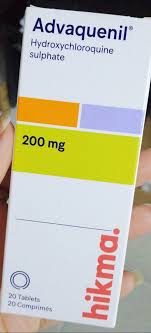
- Hydroxychloroquine: Sometimes used in mild cases.
- Cyclophosphamide: Reserved for severe, refractory cases.
- Supportive Care: Iron supplementation is critical to treat anemia. In severe acute hemorrhage, blood transfusions and mechanical ventilation may be required.
Long-term Management and Lifestyle
- Monitoring: Serial pulmonary function tests (PFTs) and HRCT scans to monitor for the development of pulmonary fibrosis.
- Nutrition: High-caloric intake is necessary for children suffering from failure to thrive.
- Avoidance: Patients should avoid known environmental triggers, including cigarette smoke and identified mold-infested environments.
6. Frequently Asked Questions (FAQ)
1. Is Idiopathic Pulmonary Hemosiderosis curable?
There is no "cure" in the traditional sense, but the condition is manageable. With early diagnosis and consistent adherence to immunosuppressive therapy, many patients lead near-normal lives.
2. What is the survival rate for IPH?
The prognosis depends on the frequency and severity of bleeding episodes. While previously associated with high mortality, modern immunosuppressive therapies have significantly improved long-term survival outcomes.
3. Does IPH always cause hemoptysis?
No. In children, hemoptysis is frequently absent, which often leads to misdiagnosis as simple iron-deficiency anemia or pneumonia.
4. Can IPH be inherited?
While most cases are sporadic, there are documented instances of familial IPH, suggesting a possible genetic component in rare cases.
5. How often should I have follow-up imaging?
Follow-up frequency is determined by the patient’s clinical stability. Typically, patients on maintenance therapy undergo HRCT scans annually or if there is a change in respiratory status.
6. What is the gold standard for diagnosis?
The gold standard is the demonstration of hemosiderin-laden macrophages in the bronchoalveolar lavage (BAL) fluid, coupled with the exclusion of other systemic diseases.
7. Is surgery required for IPH?
Surgery is rarely indicated. A lung biopsy is only performed if non-invasive methods (like BAL and serology) fail to provide a definitive diagnosis.
8. Can I live a normal life with IPH?
Yes, provided the patient remains under the care of a pulmonologist and adheres to their medication regimen to prevent pulmonary fibrosis.
9. Why is iron-deficiency anemia common in IPH?
The repeated bleeding into the alveolar spaces traps blood within the lungs. This blood is not recycled by the body, leading to a chronic loss of iron stores.
10. What are the signs of a relapse?
Patients should monitor for increased fatigue, recurrence of a dry cough, unexpected shortness of breath, or the sudden appearance of blood in the sputum.
Conclusion
Idiopathic Pulmonary Hemosiderosis is a complex, multi-faceted disease that requires a multidisciplinary approach. Early clinical suspicion, particularly in cases of unexplained iron-deficiency anemia and respiratory symptoms, is vital for improving patient outcomes. By utilizing standardized diagnostic pathways and aggressive, early immunosuppressive intervention, clinicians can effectively mitigate the risk of irreversible pulmonary scarring and improve the quality of life for those affected by this rare condition.