Clinical Assessment & Protocol
Typical Presentation (HPI)
EN: Patient presents with complaints of thoracic back discomfort and visible rounding of the upper back. Symptoms are exacerbated by prolonged sitting or static postures and are relieved by active correction or recumbency. Denies radicular symptoms, neurological deficits, bowel/bladder dysfunction, or constitutional symptoms. No history of trauma or prior spinal surgery. AR: يراجع المريض بشكوى من انزعاج في الظهر الصدري وتقوس مرئي في الجزء العلوي من الظهر. تزداد الأعراض سوءاً مع الجلوس لفترات طويلة أو الوضعيات الثابتة، وتتحسن مع التصحيح النشط أو الاستلقاء. ينفي وجود أعراض جذرية، أو عجز عصبي، أو خلل في الأمعاء/المثانة، أو أعراض عامة. لا يوجد تاريخ لصدمات أو جراحات سابقة في العمود الفقري.
General Examination
EN: Physical examination reveals an exaggerated thoracic curvature that is flexible and correctable upon active extension. No fixed deformity or gibbus deformity noted. Spine is supple with full range of motion. Neurological examination is intact, with symmetric deep tendon reflexes and no focal motor or sensory deficits. Adam’s forward bend test is negative for structural scoliosis. AR: يكشف الفحص البدني عن تقوس صدري مبالغ فيه، وهو مرن وقابل للتصحيح عند التمديد النشط. لا توجد تشوهات ثابتة أو حدبة. العمود الفقري مرن مع نطاق حركة كامل. الفحص العصبي سليم، مع منعكسات وترية عميقة متناظرة وعدم وجود عجز حركي أو حسي بؤري. اختبار الانحناء للأمام (اختبار آدم) سلبي فيما يخص الجنف البنيوي.
Treatment Protocol
EN: Recommended treatment plan includes a structured physical therapy program focusing on strengthening the thoracic extensors, scapular stabilizers, and core musculature. Emphasis on postural re-education, ergonomic adjustments at the workplace, and daily stretching exercises for the pectoralis muscles. Follow-up in 3 months to assess compliance and symptomatic improvement. AR: تتضمن خطة العلاج الموصى بها برنامجاً علاجياً فيزيائياً منظماً يركز على تقوية عضلات الباسطة الصدرية، ومثبتات لوح الكتف، وعضلات الجذع. التركيز على إعادة التثقيف الوضعي، والتعديلات المريحة في مكان العمل، وتمارين التمدد اليومية لعضلات الصدر. المتابعة بعد 3 أشهر لتقييم الالتزام والتحسن في الأعراض.
Patient Education
EN: Postural kyphosis is a functional, non-structural condition caused by habitual slouching. It is reversible through consistent exercise and conscious postural correction. Avoid prolonged static postures; take frequent breaks to perform postural resets. Maintain a neutral spine alignment during daily activities. Consistency in your home exercise program is the primary determinant of long-term success. AR: الحداب الوضعي هو حالة وظيفية غير بنيوية ناتجة عن اعتياد الانحناء. يمكن عكس الحالة من خلال التمارين المستمرة والتصحيح الواعي للوضعية. تجنب الوضعيات الثابتة لفترات طويلة؛ خذ فترات راحة متكررة للقيام بتمارين تصحيح الوضعية. حافظ على محاذاة العمود الفقري في وضع محايد أثناء الأنشطة اليومية. الالتزام ببرنامج التمارين المنزلية هو العامل الأساسي للنجاح على المدى الطويل.
Systemic & Specialized Examinations
EN: Distinct radiculopathy (L4/L5/S1). Strict Cauda Equina precautions documented. AR: اعتلال عصبي جذري واضح. تم توثيق تحذيرات متلازمة ذيل الفرس بصرامة.
Orthopedic & Trauma Assessments
EN: Heavy lifting incident with spinal rotation, or insidious degenerative disc disease. AR: حادث رفع أوزان ثقيلة مع دوران للعمود الفقري، أو انزلاق غضروفي تدريجي.
EN: Antalgic gait. Exhibits a 'list' (sciatic scoliosis) away from the affected side. Difficulty with heel/toe walk. AR: مشية متألمة. يظهر ميلاً (جنف وركي) لتخفيف الضغط. صعوبة في المشي على الكعب/الأصابع.
EN: Loss of normal lumbar lordosis. Severe paraspinal muscle spasm. AR: فقدان التقوس القطني الطبيعي. تشنج عضلي شديد حول الفقرات.
EN: Straight Leg Raise (SLR): Strongly positive at 30-45°. Slump test positive. AR: اختبار رفع الساق المستقيمة (SLR): إيجابي بقوة عند 30-45 درجة.
EN: Weakness (4/5) in EHL (L5) or Plantarflexion (S1). AR: ضعف (4/5) في باسطة الإبهام (L5) أو الثني الأخمصي (S1).
EN: Hypoesthesia to pinprick over the foot dorsum (L5) or lateral border (S1). AR: نقص الإحساس للوخز على ظهر القدم (L5) أو الجانب الوحشي (S1).
EN: Achilles (S1) diminished 1+. Patellar (L4) 2+. AR: منعكس وتر أخيل ضعيف 1+. منعكس الرضفة طبيعي 2+.
EN: DP and PT pulses 2+ symmetric. AR: النبضات الطرفية طبيعية.
Clinical Guide: Postural Kyphosis (Scheuermann’s Variant and Functional Postural Deficit)
1. Comprehensive Introduction & Overview
Postural kyphosis, often clinically referred to as "roundback" or "slouching," represents a sagittal plane spinal deformity characterized by an exaggerated thoracic curvature. Unlike structural kyphosis (such as Scheuermann’s disease), postural kyphosis is defined by its flexibility. It is essentially a functional manifestation of habitual slouching, muscle imbalance, and poor ergonomic adaptation.
While often dismissed as a benign aesthetic concern, chronic postural kyphosis can lead to significant biomechanical shifts, resulting in secondary myofascial pain syndromes, respiratory capacity reduction, and potential long-term degenerative disc disease. As an orthopedic concern, it is most prevalent in adolescents and young adults, though it is increasingly observed in sedentary office-working populations.
2. Deep-Dive: Technical Specifications and Pathophysiology
The Biomechanics of the Thoracic Spine
The thoracic spine naturally exhibits a kyphotic curve ranging from 20° to 45°. Postural kyphosis is identified when this curvature exceeds the physiological norms but remains supple under forced extension (the "Adam’s Forward Bend Test" and prone hyperextension maneuvers).
Pathophysiological Mechanisms
The development of postural kyphosis is a multi-factorial process involving:
- Adaptive Shortening: The pectoralis major and minor muscles become hypertonic and shortened due to prolonged protraction of the shoulders.
- Muscular Inhibition: The rhomboids, middle/lower trapezius, and serratus anterior experience reciprocal inhibition, leading to scapular winging and loss of thoracic stabilization.
- Proprioceptive Drift: Prolonged slouching recalibrates the patient’s "internal verticality," where the brain perceives the slouched posture as the "neutral" resting state.
- Ligamentous Creep: Over time, the posterior longitudinal ligament and the supraspinous ligaments undergo viscoelastic deformation, losing their ability to provide passive tension to the spinal column.
Clinical Staging/Grading
While there is no universally rigid staging system for postural kyphosis (unlike the Sorenson criteria for Scheuermann’s), clinicians utilize the following functional grading:
| Grade | Severity | Clinical Characteristics | Correctability |
|---|---|---|---|
| I | Mild | 45°–50° Cobb Angle | Fully reversible with verbal cueing |
| II | Moderate | 50°–60° Cobb Angle | Reversible with active exercise therapy |
| III | Severe | >60° Cobb Angle | Requires bracing or intensive physical rehabilitation |
3. Extensive Clinical Indications and Presentation
Standard Presentation
Patients typically present with complaints of "upper back fatigue," "tightness between the shoulder blades," or aesthetic concerns regarding their profile. The standard clinical presentation includes:
- Forward Head Posture (FHP): Cervical protraction to compensate for the thoracic curve, often leading to tension headaches.
- Scapular Protraction: The medial borders of the scapulae lift off the rib cage.
- Lumbar Hyperlordosis: A compensatory mechanism where the lumbar spine arches excessively to maintain a horizontal gaze.
Diagnostic Testing Protocols
To confirm a diagnosis of Postural Kyphosis rather than Scheuermann’s (structural), the following diagnostic battery is employed:
- The Hyperextension Test: The patient is placed in a prone position. If the kyphotic curve significantly reduces or vanishes, the condition is confirmed as postural/functional.
- Radiographic Assessment: Standing lateral spine X-rays are the gold standard. A Cobb angle measurement is mandatory.
- Neurological Screening: Must be performed to rule out myelopathy, although this is rare in pure postural cases.
- Respiratory Function Tests (Spirometry): In severe cases, reduced Forced Vital Capacity (FVC) may be noted due to restricted chest wall expansion.
4. Risks, Side Effects, and Contraindications
Potential Risks of Untreated Postural Kyphosis
- Degenerative Disc Disease (DDD): Abnormal load distribution leads to premature wear of the thoracic intervertebral discs.
- Chronic Myofascial Pain: Persistent strain on the erector spinae muscles.
- Respiratory Diminishment: Reduced vital capacity due to restricted rib cage excursion.
- Psychosocial Impact: Self-esteem issues related to body image.
Contraindications for Treatment
- Aggressive Manual Manipulation: High-velocity, low-amplitude (HVLA) adjustments are often contraindicated in patients with underlying osteopenia or hypermobility syndromes.
- Bracing without Exercise: The use of rigid thoracolumbar orthoses (TLSO) for postural kyphosis is generally discouraged, as it promotes "muscle atrophy of the spinal stabilizers." Bracing should only be used as a temporary corrective tool, never a long-term solution.
- Over-stretching: Stretching the already lengthened/weak posterior chain muscles can exacerbate the instability.
5. Differential Diagnosis
| Condition | Key Differentiator |
|---|---|
| Scheuermann’s Disease | Fixed deformity; radiographic evidence of vertebral wedging (>5°). |
| Ankylosing Spondylitis | Morning stiffness; elevated inflammatory markers (ESR/CRP); "Bamboo spine" on X-ray. |
| Osteoporotic Fracture | Acute pain onset; age-related; wedge fractures visible on imaging. |
| Congenital Kyphosis | Developmental failure of segmentation or formation of vertebrae. |
6. FAQ Section: Expert Answers
Q1: Is postural kyphosis permanent?
A: Generally, no. Because it is functional and not structural, it is highly responsive to corrective exercise and postural re-education.
Q2: Will a back brace fix my posture?
A: Braces provide temporary relief but can weaken the muscles responsible for holding you upright. They are not a substitute for active strengthening.
Q3: Can yoga help with postural kyphosis?
A: Yes, specifically extension-based poses (e.g., Cobra, Sphinx, or Cat-Cow). However, avoid excessive flexion-based poses that may increase the thoracic curve.
Q4: At what age is it too late to correct postural kyphosis?
A: It is never "too late." While plasticity is higher in adolescents, adults can achieve significant improvements in thoracic mobility and postural alignment through consistent physical therapy.
Q5: How long does it take to see results from physical therapy?
A: Most patients notice symptomatic relief within 2–4 weeks, while structural postural changes typically require 3–6 months of consistent intervention.
Q6: What is the "Cobb Angle" and why does it matter?
A: The Cobb angle is the standard measurement used to quantify the severity of spinal curvature on an X-ray. It dictates the treatment pathway.
Q7: Is surgery ever required for postural kyphosis?
A: Surgery is almost never indicated for postural kyphosis. It is reserved for severe structural deformities or neurological compromise.
Q8: Does sleeping position affect postural kyphosis?
A: Yes. Sleeping on the stomach can exacerbate neck and thoracic strain. A side-sleeping position with a pillow between the knees is generally recommended.
Q9: Can postural kyphosis cause breathing problems?
A: In severe cases, the rounded spine limits the expansion of the rib cage, which can lead to a slight decrease in lung capacity.
Q10: Are there specific supplements I should take?
A: While no supplement cures kyphosis, ensuring adequate Vitamin D and Calcium intake is essential for maintaining overall bone health and preventing secondary degenerative changes.
7. Clinical Management Strategy: The Three-Pillar Approach
Effective management of postural kyphosis relies on a structured, three-pillar intervention model:
Pillar 1: Myofascial Release
The goal is to reduce the hypertonicity of the anterior chain.
* Pectoral Release: Foam rolling or lacrosse ball massage to the pectoralis major/minor.
* Upper Trapezius Stretching: To alleviate tension headaches associated with the forward head posture.
Pillar 2: Neuromuscular Re-education
The goal is to change the patient’s perception of "neutral."
* Wall Angels: A critical exercise to train thoracic extension and scapular retraction simultaneously.
* Chin Tucks: To address the cervical component of the kyphotic posture.
Pillar 3: Strength and Stabilization
The goal is to build endurance in the posterior chain.
* Prone Y-T-W Raises: High-repetition, low-weight exercises to target the mid-trapezius and rhomboids.
* Thoracic Extension Mobility: Utilizing a foam roller to move the thoracic spine into extension, countering the daily "flexion bias" of modern life.
8. Conclusion
Postural kyphosis is a manageable, non-structural condition that serves as a primary marker for musculoskeletal imbalance. By focusing on the interplay between anterior tightness and posterior weakness, clinicians can effectively guide patients toward long-term spinal health. Early intervention through physical therapy and ergonomic optimization remains the gold standard, preventing the transition from a functional deficit to a permanent structural deformity.
Disclaimer: This guide is intended for clinical education purposes. Always correlate physical findings with diagnostic imaging and consult with a licensed orthopedic specialist before initiating treatment protocols.
Related Clinical Integration
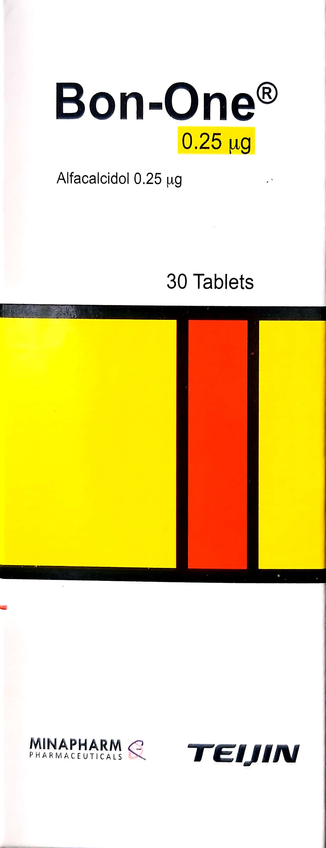
In a modern clinical setting, the management of postural kyphosis requires a comprehensive approach that bridges conservative medical therapy with advanced surgical evaluation. While postural kyphosis is primarily addressed through physical therapy and ergonomic correction, clinicians must remain vigilant for underlying metabolic or structural pathologies; in cases where bone density or mineral metabolism is a concern, pharmacological support with Calcium Gluconate / غلوكونات الكالسيوم 10ml and Bon-one / بون-ون 0.25mcg may be indicated to optimize skeletal health. For patients presenting with progressive deformity or secondary complications, surgical consultation is essential, necessitating a thorough understanding of procedures such as Cervical Spinal Fusion (ACDF) / دمج الفقرات العنقية (بالطريق الأمامي مع استئصال القرص) (عملية كبرى في غرف العمليات) and Distal Femoral Osteotomy (DFO) / قطع العظم الفخذي القاصي (عملية كبرى في غرف العمليات) when multi-level spinal or lower-limb alignment is required. Clinicians are encouraged to deepen their expertise through specialized resources, including Kyphosis & Scheuermann Disease: Comprehensive Operative Management, Unusual Causes of Kyphosis: Comprehensive Surgical Management, and Spinal Stability: Protecting Against Neurologic Deficit, while utilizing [Orthopedic Board Review MCQs: Spine Surgery Questions | Part 27](https://www.hutaifortho.com/en/hub/orthopedic-board-review-oite-aaos-part-131/orthopedic-board-review-oite-aaos-part-2