Clinical Assessment & Protocol
Typical Presentation (HPI)
EN: Patient presents with pleuritic chest pain, sharp and localized, exacerbated by deep inspiration and coughing. Associated with dyspnea, non-productive cough, and systemic symptoms of SLE flare (fatigue, arthralgia, fever). Denies hemoptysis or orthopnea. Symptoms consistent with serositis secondary to systemic lupus erythematosus. AR: يعاني المريض من ألم صدري جنبي (pleuritic)، حاد وموضعي، يزداد سوءاً مع الشهيق العميق والسعال. يترافق مع ضيق في التنفس، سعال جاف، وأعراض جهازية لنشاط الذئبة الحمراء (إرهاق، ألم مفصلي، حمى). ينفي وجود نفث دموي أو ضيق تنفس عند الاستلقاء. الأعراض تتوافق مع التهاب الأغشية المصلية الثانوي للذئبة الحمراء الجهازية.
General Examination
EN: Respiratory exam reveals decreased chest wall excursion on the affected side, dullness to percussion at the lung base, and diminished breath sounds. A pleural friction rub is audible on auscultation. No signs of tension pneumothorax or severe respiratory distress. Cardiovascular exam: S1/S2 normal, no signs of pericardial effusion or tamponade. AR: يكشف الفحص التنفسي عن انخفاض في حركة جدار الصدر في الجانب المصاب، خفوت في الأصوات عند القرع في قاعدة الرئة، وتناقص في أصوات التنفس. يُسمع احتكاك جنبي (pleural friction rub) عند التسمع. لا توجد علامات استرواح صدري توتري أو ضيق تنفس حاد. فحص القلب: الأصوات القلبية S1/S2 طبيعية، لا توجد علامات انصباب تاموري أو اندحاس قلبي.
Treatment Protocol
EN: Initiate NSAIDs for symptomatic relief of pleuritic pain. Consider short-course systemic corticosteroids (e.g., Prednisone) for moderate-to-severe serositis. Optimize baseline SLE therapy (Hydroxychloroquine, immunosuppressants). Monitor for pleural effusion; perform thoracentesis if clinically indicated for diagnostic or therapeutic purposes. AR: البدء بمضادات الالتهاب غير الستيرويدية (NSAIDs) لتخفيف ألم الصدر الجنبِي. النظر في استخدام دورة قصيرة من الكورتيكوستيرويدات الجهازية (مثل بريدنيزون) في حالات التهاب الأغشية المصلية المتوسطة إلى الشديدة. تحسين العلاج الأساسي للذئبة (هيدروكسي كلوروكوين، مثبطات المناعة). المراقبة تحسباً لحدوث انصباب جنبي؛ إجراء بزل الصدر (thoracentesis) إذا استدعت الضرورة السريرية لأغراض تشخيصية أو علاجية.
Patient Education
EN: Lupus pleuritis is an inflammation of the lung lining caused by your underlying autoimmune condition. Take medications as prescribed to control systemic inflammation. Seek immediate medical attention if you experience worsening shortness of breath, high fever, or chest pain that prevents you from breathing deeply. Maintain regular follow-ups to monitor SLE activity. AR: التهاب غشاء الجنب المرتبط بالذئبة هو التهاب في بطانة الرئة ناتج عن حالتك المناعية الذاتية. التزم بتناول الأدوية الموصوفة للسيطرة على الالتهاب الجهازي. اطلب الرعاية الطبية الفورية إذا شعرت بزيادة في ضيق التنفس، أو ارتفاع في درجة الحرارة، أو ألم صدري يمنعك من التنفس بعمق. حافظ على المتابعة الدورية لمراقبة نشاط مرض الذئبة الحمراء.
Systemic & Specialized Examinations
EN: S1, S2 present. No murmurs. Normal rate and rhythm. AR: صوتا القلب الأول والثاني طبيعيان. لا توجد نفخات.
EN: Chest examination reveals [decreased breath sounds/friction rub] at the [location]. Percussion note is [dull/resonant]. Oxygen saturation is [percentage] on room air. AR: يكشف فحص الصدر عن [انخفاض في أصوات التنفس/احتكاك جنبي] في [الموقع]. صوت القرع هو [مكتوم/رنان]. تشبع الأكسجين هو [النسبة المئوية] في هواء الغرفة.
EN: Abdomen soft, non-tender, non-distended. AR: البطن لين ولا يوجد ألم.
EN: Alert, oriented x3. No focal deficits. AR: المريض واعي ومدرك. لا يوجد عجز عصبي بؤري.
EN: Unremarkable or not routinely indicated for this specific respiratory pathology. AR: طبيعي أو غير مطلوب روتينياً لهذا المرض التنفسي.
EN: Unremarkable or not routinely indicated for this specific respiratory pathology. AR: طبيعي أو غير مطلوب روتينياً لهذا المرض التنفسي.
EN: Unremarkable or not routinely indicated for this specific respiratory pathology. AR: طبيعي أو غير مطلوب روتينياً لهذا المرض التنفسي.
EN: Unremarkable or not routinely indicated for this specific respiratory pathology. AR: طبيعي أو غير مطلوب روتينياً لهذا المرض التنفسي.
EN: Unremarkable or not routinely indicated for this specific respiratory pathology. AR: طبيعي أو غير مطلوب روتينياً لهذا المرض التنفسي.
Orthopedic & Trauma Assessments
EN: Unremarkable or not routinely indicated for this specific respiratory pathology. AR: طبيعي أو غير مطلوب روتينياً لهذا المرض التنفسي.
EN: Unremarkable or not routinely indicated for this specific respiratory pathology. AR: طبيعي أو غير مطلوب روتينياً لهذا المرض التنفسي.
EN: Unremarkable or not routinely indicated for this specific respiratory pathology. AR: طبيعي أو غير مطلوب روتينياً لهذا المرض التنفسي.
EN: Unremarkable or not routinely indicated for this specific respiratory pathology. AR: طبيعي أو غير مطلوب روتينياً لهذا المرض التنفسي.
EN: Unremarkable or not routinely indicated for this specific respiratory pathology. AR: طبيعي أو غير مطلوب روتينياً لهذا المرض التنفسي.
EN: Unremarkable or not routinely indicated for this specific respiratory pathology. AR: طبيعي أو غير مطلوب روتينياً لهذا المرض التنفسي.
EN: Unremarkable or not routinely indicated for this specific respiratory pathology. AR: طبيعي أو غير مطلوب روتينياً لهذا المرض التنفسي.
EN: Unremarkable or not routinely indicated for this specific respiratory pathology. AR: طبيعي أو غير مطلوب روتينياً لهذا المرض التنفسي.
EN: Unremarkable or not routinely indicated for this specific respiratory pathology. AR: طبيعي أو غير مطلوب روتينياً لهذا المرض التنفسي.
1. Executive Overview: Understanding Lupus Pleuritis
Lupus Pleuritis, clinically classified under ICD-10 code M32.13, is a significant pulmonary manifestation of Systemic Lupus Erythematosus (SLE). It is characterized by the inflammation of the pleura—the double-layered membrane that surrounds the lungs and lines the chest cavity.
In patients with SLE, the immune system mistakenly attacks healthy tissues, leading to systemic inflammation. When this process targets the pleural space, it results in pleuritis, which is the most frequent thoracic involvement in lupus, occurring in approximately 40% to 60% of patients throughout the course of their disease. While often manageable, it can lead to pleural effusions—the accumulation of excess fluid—which can severely compromise respiratory function if left untreated. This guide provides a clinical overview of the pathophysiology, diagnostic pathways, and therapeutic standards for managing this condition.
2. Pathophysiology, Etiology, and Risk Factors
The Pathophysiological Mechanism
The development of Lupus Pleuritis is rooted in the fundamental autoimmune nature of SLE. The primary driver is the deposition of immune complexes (antigen-antibody complexes) within the pleural microvasculature.
- Autoantibody Formation: The presence of antinuclear antibodies (ANAs) and anti-dsDNA antibodies leads to the formation of circulating immune complexes.
- Complement Activation: These complexes deposit in the pleural tissue, triggering the complement cascade. This leads to the recruitment of neutrophils and macrophages to the pleural space.
- Cytokine Release: The resulting inflammatory milieu includes high levels of pro-inflammatory cytokines such as IL-6, TNF-alpha, and Interferon-gamma, which increase capillary permeability.
- Effusion Formation: Increased permeability allows protein-rich fluid to leak into the pleural space, resulting in an exudative pleural effusion.
Etiology and Risk Factors
Lupus Pleuritis is rarely an isolated event; it is almost exclusively secondary to systemic SLE. Risk factors that increase the likelihood of developing this complication include:
* Disease Duration: Patients with long-standing SLE are at higher risk.
* Serositis History: Patients who have previously experienced pericarditis or peritonitis.
* High Disease Activity: Poorly controlled SLE, indicated by high anti-dsDNA titers and low complement (C3/C4) levels.
* Genetic Predisposition: HLA-DR2 and HLA-DR3 alleles are often associated with increased susceptibility to SLE-related serositis.
3. Signs, Symptoms, and Clinical Presentation
The clinical presentation of Lupus Pleuritis can range from mild, self-limiting discomfort to acute respiratory distress.
Cardinal Symptoms
- Pleuritic Chest Pain: This is the hallmark symptom. It is described as sharp, stabbing pain that worsens during deep inspiration, coughing, or sneezing.
- Dyspnea: Shortness of breath, particularly upon exertion, often correlates with the volume of the pleural effusion.
- Non-Productive Cough: Often triggered by the irritation of the pleural lining.
- Systemic SLE Symptoms: Fever, fatigue, arthralgia, and malar rash often accompany a flare of pleuritis.
Physical Examination Findings
During a clinical examination, the following signs are typically noted:
* Pleural Friction Rub: A characteristic "leathery" or "creaking" sound heard through a stethoscope during respiration, caused by the rubbing of inflamed pleural surfaces.
* Dullness to Percussion: Indicative of fluid accumulation (effusion).
* Decreased Breath Sounds: Observed over the area of the effusion.
* Egophony: Increased resonance over the fluid-filled area.
4. Standard Diagnostic Evaluation & Workup
Diagnosing Lupus Pleuritis requires a multidisciplinary approach, combining rheumatological assessment with pulmonology diagnostics.
Diagnostic Workup Table
| Diagnostic Tool | Clinical Purpose | Expected Findings in Lupus Pleuritis |
|---|---|---|
| Chest X-Ray | Initial screening | Blunting of costophrenic angles; presence of effusion. |
| Thoracic Ultrasound | Bedside assessment | Precise localization and quantification of fluid. |
| Thoracentesis | Gold Standard for fluid analysis | Exudative fluid; low glucose; high LDH; presence of LE cells. |
| CT Chest (HRCT) | Structural evaluation | Pleural thickening, nodularity, or underlying parenchymal disease. |
| Serology | SLE confirmation | Elevated ANA, anti-dsDNA, low C3/C4 levels. |
Diagnostic Criteria (Light’s Criteria)
To classify the fluid as an exudate (typical of Lupus Pleuritis), the fluid must meet at least one of Light’s criteria:
1. Pleural fluid protein/serum protein ratio > 0.5.
2. Pleural fluid LDH/serum LDH ratio > 0.6.
3. Pleural fluid LDH > 2/3 of the upper limit of normal serum LDH.
5. Therapeutic Interventions
Management of Lupus Pleuritis focuses on controlling the systemic autoimmune process and providing symptomatic relief.
Pharmacotherapy
- Non-Steroidal Anti-Inflammatory Drugs (NSAIDs): Used for mild cases to manage pleuritic pain and inflammation. Caution is advised due to potential renal side effects in SLE patients.
- Systemic Corticosteroids: Prednisone is the standard of care for moderate to severe cases. It effectively suppresses the inflammatory response in the pleura.
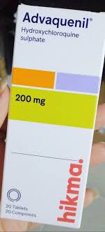
- Hydroxychloroquine: Often used as a baseline maintenance therapy to prevent future flares of serositis.
- Immunosuppressants: In refractory or recurrent cases, agents such as Azathioprine, Mycophenolate Mofetil, or Belimumab may be initiated to achieve long-term disease control.
Surgical and Procedural Interventions
- Therapeutic Thoracentesis: Indicated if the effusion is large and causing significant respiratory compromise or severe dyspnea.
- Pleurodesis: Rarely required for lupus-related effusions, but may be considered in cases of chronic, recurrent, and symptomatic effusions that do not respond to systemic immunosuppression.
Lifestyle and Long-Term Management
- Smoking Cessation: Essential to reduce further irritation of the pulmonary system.
- Monitoring: Regular pulmonary function tests (PFTs) and serial imaging for patients with a history of recurrent pleural involvement.
- Vaccination: Patients on immunosuppressants must be up-to-date on pneumococcal and influenza vaccinations.
6. Frequently Asked Questions (FAQ)
1. Is Lupus Pleuritis fatal?
Rarely. While it can cause significant discomfort and respiratory distress, it is usually managed effectively with corticosteroids and disease-modifying anti-rheumatic drugs (DMARDs).
2. How do I differentiate between Lupus Pleuritis and pneumonia?
Pneumonia is typically infectious and characterized by productive cough and fever. Lupus Pleuritis is inflammatory, often accompanied by other systemic lupus symptoms like joint pain or rashes, and the pleural fluid analysis will show an exudative profile without bacteria.
3. Will the fluid in my lungs go away on its own?
Small effusions may resolve as the systemic lupus flare subsides. However, moderate to large effusions usually require medical intervention to prevent respiratory distress.
4. Can I exercise with Lupus Pleuritis?
During an acute flare, physical activity should be limited to prevent worsening of pleuritic pain and oxygen demand. Once the inflammation is controlled, light exercise is encouraged.
5. How often do I need a chest X-ray?
Your rheumatologist or pulmonologist will determine this based on your symptoms. In stable patients, routine screening may not be necessary, but it is standard during suspected flares.
6. Does Lupus Pleuritis lead to permanent lung damage?
In most cases, it does not. However, chronic, recurrent inflammation can lead to pleural thickening or fibrosis, which may result in restrictive lung disease over time.
7. Are there specific diets to help with pleural inflammation?
While no specific diet cures lupus, an anti-inflammatory diet rich in Omega-3 fatty acids and antioxidants may support overall systemic health.
8. Is pleuritic chest pain always a sign of a lupus flare?
While it is a common sign of a flare, chest pain can also indicate other conditions like pulmonary embolism or pericarditis. You must seek medical evaluation to rule out life-threatening causes.
9. Can pregnancy affect Lupus Pleuritis?
Pregnancy can alter immune function and may increase the risk of lupus flares. Patients planning pregnancy should be closely monitored by a high-risk obstetrician and rheumatologist.
10. What is the role of Hydroxychloroquine in managing this?
Hydroxychloroquine is the cornerstone of SLE maintenance therapy. It has been shown to reduce the frequency of disease flares, including serositis like pleuritis, and improves long-term survival.