Clinical Assessment & Protocol
Typical Presentation (HPI)
History of frequent fractures and delayed dental eruption; risk of osteomyelitis after extractions.
General Examination
Unremarkable or not routinely indicated.
Treatment Protocol
Focus on infection prevention; atraumatic surgical procedures.
Patient Education
High risk of osteomyelitis; avoid unnecessary extractions.
Systemic & Specialized Examinations
EN: S1, S2 present. No murmurs. AR: صوتا القلب الأول والثاني طبيعيان. لا توجد نفخات.
EN: Lungs clear to auscultation. AR: الرئتان صافيتان عند التسمع.
EN: Abdomen soft, non-tender. AR: البطن لين ولا يوجد ألم.
EN: Alert, oriented x3. No focal deficits. AR: المريض واعي ومدرك. لا يوجد عجز عصبي بؤري.
EN: Unremarkable or not routinely indicated. AR: طبيعي أو غير مطلوب روتينياً.
EN: Unremarkable or not routinely indicated. AR: طبيعي أو غير مطلوب روتينياً.
EN: Unremarkable or not routinely indicated. AR: طبيعي أو غير مطلوب روتينياً.
EN: Unremarkable or not routinely indicated. AR: طبيعي أو غير مطلوب روتينياً.
EN: Dense bone appearance on radiographs; narrow pulp chambers; increased risk of jaw necrosis. AR: مظهر عظمي كثيف في الأشعة؛ حجرات لبية ضيقة؛ خطر متزايد لنخر الفك.
1. Comprehensive Introduction & Overview
Osteopetrosis, historically referred to as "marble bone disease" or Albers-Schönberg disease, is a rare, heterogeneous group of hereditary disorders characterized by a fundamental defect in osteoclast function. Unlike osteoporosis, where bone density is reduced, osteopetrosis is defined by a pathological increase in bone mass and density due to the failure of bone resorption.
The skeletal system is a dynamic organ that undergoes constant remodeling. This process requires a delicate balance between osteoblasts (bone-forming cells) and osteoclasts (bone-resorbing cells). In patients with osteopetrosis, the osteoclasts are either absent, dysfunctional, or incapable of acidifying the resorption lacunae, leading to the accumulation of dense, brittle, and structurally disorganized bone.
Clinical Classification
Osteopetrosis is broadly classified into two primary clinical phenotypes based on the mode of inheritance and the severity of clinical presentation:
| Type | Inheritance | Severity | Clinical Focus |
|---|---|---|---|
| Autosomal Recessive (ARO) | Recessive | Severe | Infantile, often fatal early, hematologic failure |
| Autosomal Dominant (ADO) | Dominant | Mild to Moderate | Benign, adult-onset, often asymptomatic |
2. Deep-Dive into Pathophysiology
The pathophysiology of osteopetrosis centers on the failure of the osteoclast to perform its primary function: the resorption of mineralized bone.
The Osteoclast Mechanism
Under normal physiological conditions, osteoclasts attach to the bone surface, creating a "sealing zone" via integrins. They then secrete hydrogen ions (via V-ATPase pumps) and lysosomal enzymes (such as Cathepsin K) into the resorption pit to dissolve the hydroxyapatite mineral and the collagenous matrix.
In osteopetrosis, this mechanism is disrupted. The underlying genetic mutations often affect:
1. Proton Pump Function: Mutations in the TCIRG1 gene prevent the acidification of the resorption pit.
2. Chloride Channel Function: Mutations in CLCN7 prevent the efflux of chloride ions, which is necessary to maintain electrical neutrality during proton transport.
3. RANK/RANKL Signaling: Disruptions in the RANK-RANKL-OPG signaling pathway prevent the proper differentiation and maturation of osteoclast precursors.
Consequences of Pathological Bone Density
The accumulation of primary spongiosa (unresorbed cartilage and bone) leads to:
* Medullary Canal Obliteration: The marrow space is encroached upon, leading to myelophthisic anemia, thrombocytopenia, and leukopenia.
* Cranial Nerve Compression: The narrowing of the skull foramina leads to pressure on cranial nerves, specifically the optic (II), oculomotor (III), facial (VII), and vestibulocochlear (VIII) nerves.
* Mechanical Brittleness: Despite the increased density, the bone is disorganized and lacks proper trabecular orientation, leading to a paradoxical increase in fracture risk (the "chalk-stick" fracture).
3. Clinical Indications & Standard Presentation
The clinical presentation varies significantly based on the genetic subtype and the age of onset.
Infantile Malignant Osteopetrosis (ARO)
Usually presents within the first year of life.
* Hematologic: Severe anemia, extramedullary hematopoiesis (splenomegaly and hepatomegaly).
* Neurologic: Optic nerve atrophy (leading to blindness), facial nerve palsy, and hearing loss.
* Metabolic: Hypocalcemia and secondary hyperparathyroidism.
* Growth: Failure to thrive, short stature.
Adult-Onset Osteopetrosis (ADO)
Often diagnosed incidentally during routine radiographic examination.
* Skeletal: Recurrent fractures (often low-energy), osteomyelitis (frequently of the mandible, especially after dental extraction).
* Neurologic: Progressive hearing loss (due to bone overgrowth in the internal auditory canal) and cranial nerve palsies.
* Hematologic: Usually mild or absent.
4. Diagnostic Testing & Imaging
The diagnosis of osteopetrosis relies on a combination of clinical suspicion, radiographic findings, and genetic confirmation.
Radiographic Findings
- Generalized Sclerosis: Increased bone density (radiopacity) on plain films.
- "Bone-within-a-Bone" (Endobone): A pathognomonic appearance where the shape of the bone at an earlier age is visible within the adult bone.
- "Sandwich Vertebrae": Dense bands at the superior and inferior endplates of the vertebrae.
- Erlenmeyer Flask Deformity: Failure of the metaphysis to remodel, leading to a widened, flask-like shape in long bones.
Laboratory Markers
- Complete Blood Count (CBC): To assess for anemia and pancytopenia.
- Calcium/Phosphate/PTH: To differentiate from rickets or hyperparathyroidism.
- Acid Phosphatase (TRAP): Elevated levels of Tartrate-Resistant Acid Phosphatase are often found, reflecting the high number of dysfunctional osteoclasts.
Genetic Testing
Molecular analysis is the gold standard for diagnosis. Targeted gene panels covering TCIRG1, CLCN7, OSTM1, SNX10, and RANKL are used to confirm the mutation and facilitate genetic counseling.
5. Risks, Side Effects, and Management
Management is largely supportive, as there is no cure for the underlying genetic defect.
Standard Management Protocols
- Hematopoietic Stem Cell Transplantation (HSCT): This is the only curative therapy for the severe infantile form (ARO). It replaces defective osteoclast precursors with healthy donor cells, allowing for normal bone remodeling.
- Medical Therapy:
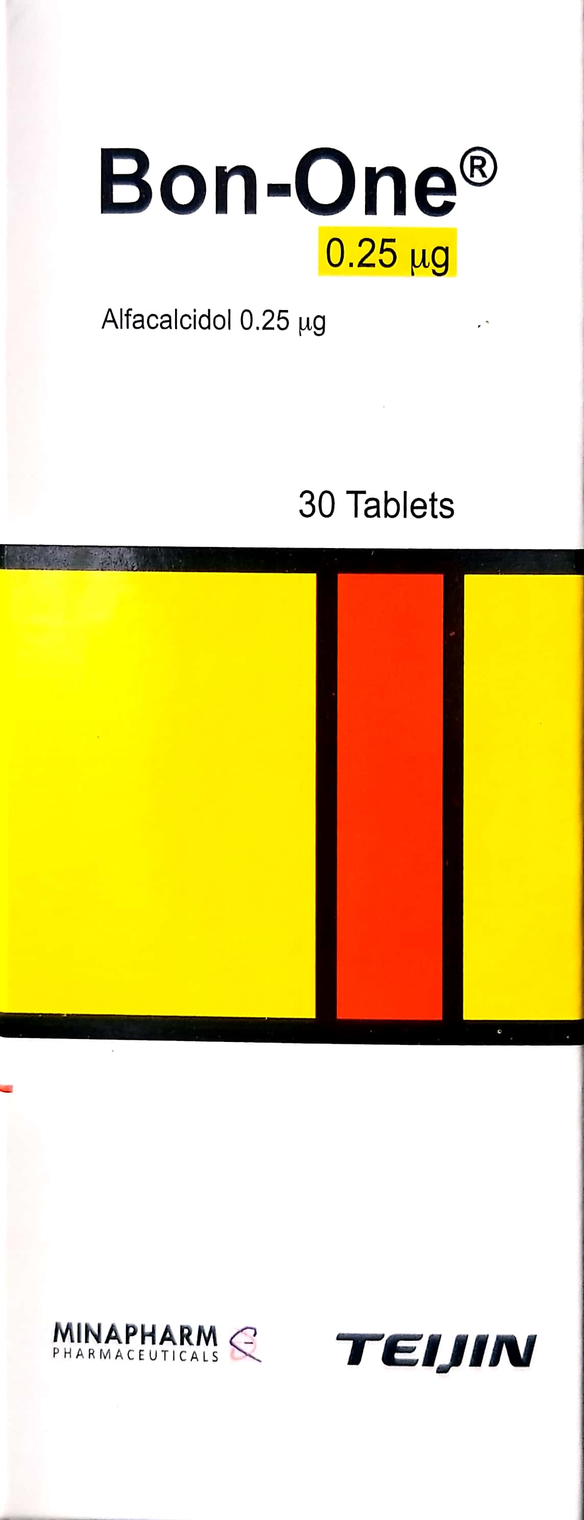
- Calcitriol: Used to stimulate bone resorption and improve calcium balance.
- Interferon Gamma-1b: Used to improve leukocyte function and potentially osteoclast activity.
- Erythropoietin: To manage anemia.
- Surgical Intervention: Orthopedic management of fractures (intramedullary nailing) and decompression surgery for cranial nerve entrapment.
Contraindications & Risks
- Bisphosphonates: Absolutely contraindicated, as they further inhibit osteoclast function.
- Dental Care: Patients are at high risk of osteomyelitis of the jaw following dental procedures. Prophylactic antibiotics and meticulous oral hygiene are mandatory.
- High-Impact Sports: Due to the brittle nature of the bone, patients should avoid contact sports.
6. Massive FAQ Section
Q1: Is osteopetrosis the same as osteoporosis?
No. Osteoporosis is a decrease in bone mass and density, leading to porous bones. Osteopetrosis is the opposite—an increase in bone mass and density, leading to brittle, disorganized bones.
Q2: Is osteopetrosis hereditary?
Yes. It is a genetic disorder. Depending on the mutation, it can be inherited in an autosomal recessive (severe) or autosomal dominant (mild) pattern.
Q3: What is the "bone-within-a-bone" sign?
This is a classic radiological feature where the dense silhouette of a smaller bone is visible inside the larger, mature bone, reflecting a period of arrested bone resorption.
Q4: Why do patients with osteopetrosis get anemia?
The dense bone obliterates the marrow space where blood cells are produced (hematopoiesis). This forces the body to produce blood cells in the liver and spleen (extramedullary hematopoiesis), leading to organ enlargement.
Q5: Can osteopetrosis be cured?
For the severe infantile form, hematopoietic stem cell transplantation (HSCT) can be curative if performed early. For the adult-onset form, treatment is supportive.
Q6: Why are dental extractions dangerous for these patients?
The bone in osteopetrosis has poor vascularity. Following an extraction, the bone is highly susceptible to osteomyelitis (bone infection) because it cannot heal properly.
Q7: What is the "Erlenmeyer flask deformity"?
It is a widening of the metaphysis of long bones (typically seen in the distal femur) caused by a failure of the bone to remodel its shape during growth.
Q8: Does everyone with osteopetrosis have the same symptoms?
No. The severity varies wildly from individuals who are completely asymptomatic and diagnosed by accident, to infants with life-threatening hematologic and neurologic complications.
Q9: What is the role of the osteoclast in this disease?
The osteoclast is the "villain" of the story. In osteopetrosis, the osteoclast fails to break down old bone. This means that instead of being replaced by new, healthy bone, the old bone stays, becomes overly dense, and loses its structural integrity.
Q10: Are there any specific diet recommendations?
While diet cannot cure the disease, patients are often monitored for calcium and Vitamin D levels to ensure they do not develop secondary hyperparathyroidism or rickets, which can complicate the skeletal fragility.
7. Prognosis and Long-Term Outlook
The prognosis for osteopetrosis is highly dependent on the subtype.
- Infantile (ARO): Without successful HSCT, the prognosis is poor, with many patients succumbing to infection, hemorrhage, or severe neurologic failure within the first decade of life.
- Adult (ADO): The prognosis is generally good. Life expectancy is often normal, though patients must manage recurrent fractures, dental infections, and potential sensory loss (hearing/vision).
Conclusion for Clinical Practitioners
Osteopetrosis represents a fascinating intersection of hematology, genetics, and orthopedics. For the clinician, the priority is early recognition of the radiological signs, careful monitoring of hematologic status, and the prevention of secondary complications like osteomyelitis. As genetic therapies continue to evolve, the outlook for patients—particularly those with the severe infantile form—is gradually improving, shifting from palliative care to potential long-term management and cure.