Clinical Assessment & Protocol
Typical Presentation (HPI)
Bone pain, muscle cramps, and dental sensitivity.
General Examination
Unremarkable or not routinely indicated.
Treatment Protocol
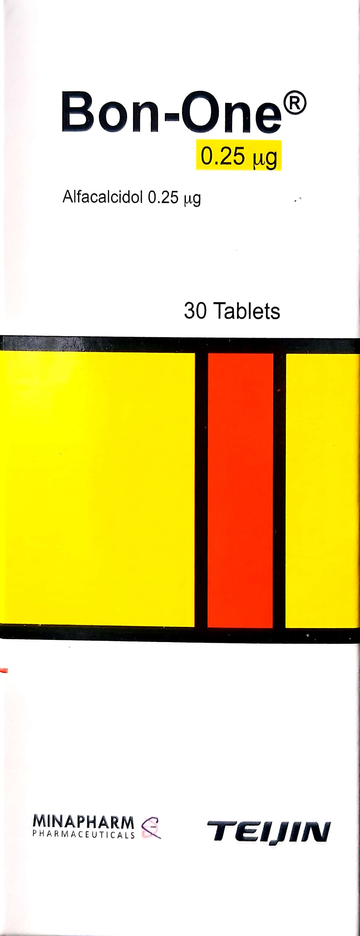
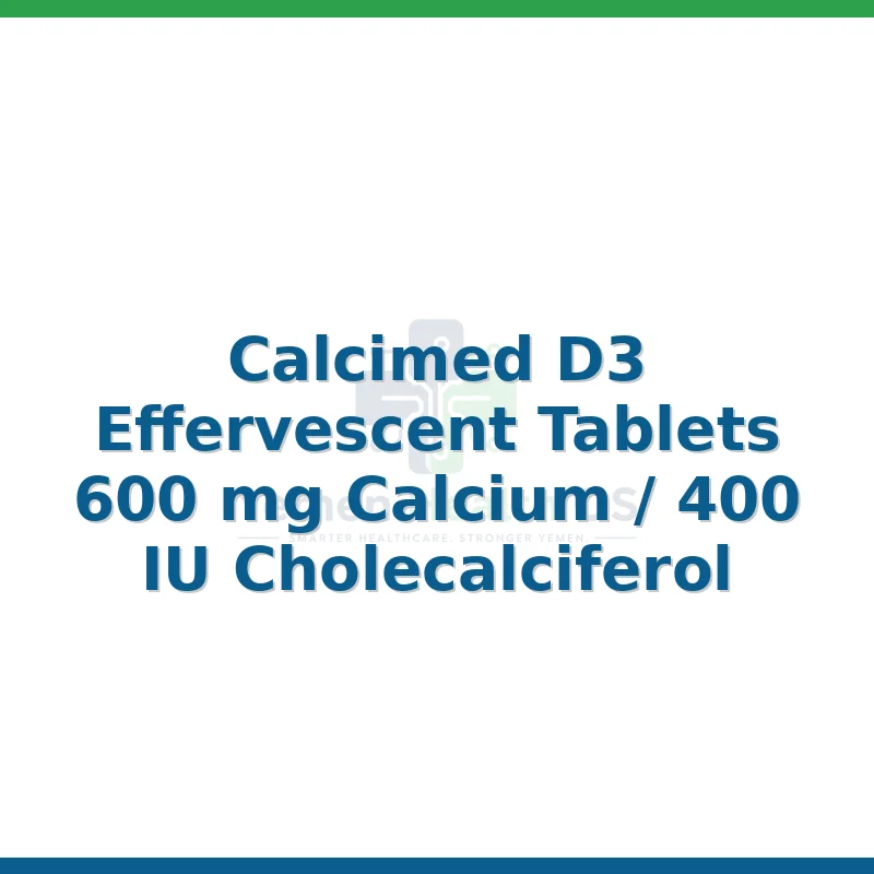
High-dose Calcium citrate and Vitamin D3 supplementation.
Patient Education
Calcium citrate is preferred over carbonate due to gastric acid independence.
Systemic & Specialized Examinations
EN: S1, S2 present. No murmurs. AR: صوتا القلب الأول والثاني طبيعيان. لا توجد نفخات.
EN: Lungs clear to auscultation. AR: الرئتان صافيتان عند التسمع.
EN: Positive Chvostek or Trousseau signs in severe cases. AR: علامات شوفستيك أو تروسو إيجابية في الحالات الشديدة.
EN: Alert, oriented x3. No focal deficits. AR: المريض واعي ومدرك. لا يوجد عجز عصبي بؤري.
EN: Unremarkable or not routinely indicated. AR: طبيعي أو غير مطلوب روتينياً.
EN: Unremarkable or not routinely indicated. AR: طبيعي أو غير مطلوب روتينياً.
EN: Unremarkable or not routinely indicated. AR: طبيعي أو غير مطلوب روتينياً.
EN: Unremarkable or not routinely indicated. AR: طبيعي أو غير مطلوب روتينياً.
EN: Unremarkable or not routinely indicated. AR: طبيعي أو غير مطلوب روتينياً.
1. Comprehensive Introduction & Overview
Post-Bariatric Hypocalcemia (PBH), often manifesting as secondary hyperparathyroidism (SHPT), represents one of the most critical long-term metabolic complications following bariatric surgical interventions. As the prevalence of metabolic and bariatric surgery (MBS)—specifically Roux-en-Y gastric bypass (RYGB) and biliopancreatic diversion with duodenal switch (BPD/DS)—continues to rise, clinicians must maintain a high index of suspicion for calcium-phosphorus-parathyroid hormone (PTH) axis dysregulation.
At its core, this condition is a state of chronic calcium deficiency induced by a combination of reduced gastric acid production, bypassed absorptive surface area, and poor adherence to micronutrient supplementation. When serum calcium levels drop, the parathyroid glands respond by over-secreting PTH to mobilize calcium from the skeletal system. This compensatory mechanism, while initially protective of serum calcium, leads to deleterious effects on bone mineral density (BMD), increasing the risk of osteopenia, osteoporosis, and fragility fractures.
2. Deep-Dive: Etiology and Pathophysiology
The pathophysiology of PBH is multifactorial and rooted in the physiological changes mandated by modern bariatric procedures.
The Mechanism of Malabsorption
- Achlorhydria and Hypochlorhydria: Calcium carbonate, the most common form of supplemental calcium, requires an acidic environment for optimal ionization and absorption. Bariatric procedures significantly reduce gastric acid secretion, rendering calcium carbonate largely insoluble and poorly absorbed.
- Anatomical Bypassing: RYGB and BPD/DS bypass the duodenum and proximal jejunum, which are the primary sites for active, vitamin D-dependent calcium absorption.
- Vitamin D Deficiency: Vitamin D is fat-soluble. The malabsorptive nature of these surgeries impairs the absorption of dietary fats and fat-soluble vitamins, leading to profound secondary deficiency, which further exacerbates the inability to absorb calcium.
The PTH Axis Feedback Loop
The metabolic cascade follows a predictable trajectory:
* Step 1: Decreased calcium absorption leads to a decline in ionized serum calcium.
* Step 2: The calcium-sensing receptors (CaSR) on the parathyroid glands detect the drop.
* Step 3: PTH secretion increases (Secondary Hyperparathyroidism).
* Step 4: PTH stimulates osteoclast activity to release calcium from the bone matrix.
* Step 5: Chronic bone resorption leads to a net loss of bone mass, predisposing the patient to skeletal fragility.
| Factor | Impact on Calcium Homeostasis |
|---|---|
| Gastric Acid Reduction | Inhibits ionization of calcium carbonate |
| Duodenal Bypass | Bypasses primary active transport site |
| Fat Malabsorption | Limits Vitamin D uptake |
| PTH Elevation | Chronic resorption of bone matrix |
3. Clinical Indications, Presentation, and Staging
Clinical Presentation
PBH is frequently insidious. Patients may remain asymptomatic for years while the "silent" skeletal damage progresses. When symptoms do manifest, they typically include:
* Neuromuscular: Perioral numbness, paresthesia in extremities, muscle cramps, and in severe cases, tetany or Chvostek’s/Trousseau’s signs.
* Skeletal: Diffuse bone pain, back pain, and recurring stress fractures.
* Cardiac: Potential for QT-interval prolongation on ECG.
Staging of Post-Bariatric Mineral Metabolism
The following grading system serves as a clinical framework for assessing severity:
| Stage | Serum Calcium | Serum PTH | Serum 25(OH)D | Clinical Status |
|---|---|---|---|---|
| Stage 0 | Normal | Normal | Normal | Normal Homeostasis |
| Stage 1 | Low-Normal | Elevated | Low | Subclinical SHPT |
| Stage 2 | Low | High | Very Low | Overt SHPT/Osteopenia |
| Stage 3 | Symptomatic | Markedly High | Critically Low | Bone disease/Tetany |
4. Diagnostic Evaluation and Differential Diagnosis
Key Diagnostic Tests
A comprehensive metabolic workup for a post-bariatric patient suspected of hypocalcemia must include:
1. Serum Calcium (Albumin-adjusted): Essential to avoid false negatives in patients with hypoalbuminemia.
2. Intact PTH (iPTH): The gold standard for identifying SHPT.
3. 25-Hydroxyvitamin D (25(OH)D): To assess stores; target levels should ideally be >30 ng/mL.
4. 24-Hour Urine Calcium: Critical to rule out hypercalciuria (which can occur if supplementation is excessive) and to assess intestinal absorption efficiency.
5. DEXA Scan: Baseline and periodic dual-energy X-ray absorptiometry to monitor BMD.
Differential Diagnosis
Clinicians must distinguish PBH from:
* Primary Hyperparathyroidism: Usually characterized by high calcium AND high PTH.
* Vitamin D Resistance: Rare, but requires exclusion if supplementation fails.
* Chronic Kidney Disease (CKD): A common confounder; renal function (eGFR) must be assessed to rule out renal osteodystrophy.
5. Management and Long-Term Prognosis
Therapeutic Strategy
The cornerstone of management is aggressive, tailored supplementation:
* Calcium Citrate: Preferred over Calcium Carbonate due to its solubility independent of gastric pH.
* Vitamin D3 (Cholecalciferol): Often required in high doses (e.g., 3,000–5,000 IU daily) to maintain sufficiency.
* Monitoring: Quarterly biochemical assessment in the first year, transitioning to annual screenings thereafter.
Long-Term Prognosis
If managed early, the prognosis is excellent, and skeletal recovery is possible. However, if left unchecked, the prognosis involves a significantly elevated risk of secondary osteoporosis and low-impact fractures. Patient compliance with lifelong micronutrient protocols is the single greatest determinant of long-term skeletal health.
6. Risks, Side Effects, and Contraindications
- Hypercalciuria/Nephrolithiasis: Excessive calcium supplementation, while intended to solve hypocalcemia, can lead to kidney stones. Monitoring 24-hour urine calcium is vital.
- Gastrointestinal Distress: High doses of calcium salts can cause constipation and bloating, leading to poor patient adherence.
- Contraindications: Patients with a history of hypercalcemic disorders or sarcoidosis should be managed with extreme caution regarding Vitamin D and calcium loading.
7. Massive FAQ Section (10 Critical Questions)
Q1: Why is Calcium Citrate better than Calcium Carbonate for bariatric patients?
Calcium citrate does not require gastric acid for absorption. Since bariatric surgery reduces stomach acid, Calcium Carbonate often passes through the GI tract without being absorbed, whereas Citrate is readily bioavailable.
Q2: What is the target level for Vitamin D in these patients?
Most guidelines recommend maintaining serum 25(OH)D levels above 30 ng/mL (75 nmol/L), though some specialists argue for higher targets (40–50 ng/mL) for patients with high fracture risk.
Q3: How often should I check PTH levels post-surgery?
For the first year, every 3 months is standard. Once stability is achieved and the patient is on a maintenance supplement regimen, annual checks are usually sufficient.
Q4: Can secondary hyperparathyroidism be reversed?
Yes, in most cases, normalizing calcium and Vitamin D levels will cause PTH levels to return to the normal range, effectively resolving the secondary hyperparathyroidism.
Q5: Does the type of bariatric surgery change the risk?
Yes. Malabsorptive procedures like BPD/DS carry a significantly higher risk of calcium and vitamin deficiency compared to restrictive procedures like the gastric sleeve (VSG).
Q6: What are the early signs of hypocalcemia?
Look for "numbness and tingling" (paresthesia) around the mouth, fingers, or toes, and unexplained muscle twitching or cramping.
Q7: Are there interactions with other medications?
Yes. Calcium can interfere with the absorption of levothyroxine, iron, and certain antibiotics (e.g., tetracyclines). Patients should space these medications at least 2–4 hours apart from calcium supplements.
Q8: What if my patient is allergic to calcium supplements?
This is rare. If the patient experiences severe GI side effects, consider splitting the doses throughout the day or switching to a liquid or chewable formulation.
Q9: Does bariatric surgery cause permanent bone loss?
Surgery causes an initial rapid phase of bone resorption as the body adjusts to new nutrient intake. If the patient is well-supplemented, bone turnover usually stabilizes. Without supplementation, the loss can become irreversible.
Q10: When should a DEXA scan be ordered?
A baseline DEXA scan should be performed pre-operatively or as soon as possible post-operatively, with follow-ups every 2 years, or more frequently if high turnover markers (like elevated PTH) persist.
Disclaimer: This guide is intended for clinical educational purposes and does not replace professional medical judgment. Always tailor treatment to the specific metabolic profile of the individual patient.