Clinical Assessment & Protocol
Typical Presentation (HPI)
EN: Patient presents with insidious onset of dorsal midfoot pain in the left foot, exacerbated by weight-bearing activities and high-impact loading. Reports no history of acute trauma. Pain is localized to the "N-spot" and worsens with athletic activity, improving with rest. No nocturnal pain or constitutional symptoms noted. AR: يعاني المريض من ألم تدريجي في ظهر القدم اليسرى (منطقة منتصف القدم)، يزداد سوءاً مع تحميل الوزن والأنشطة عالية التأثير. لا يوجد تاريخ لإصابة حادة. يتركز الألم في منطقة العظم الزورقي (N-spot) ويزداد مع النشاط الرياضي ويتحسن مع الراحة. لا يوجد ألم ليلي أو أعراض عامة.
General Examination
EN: Left foot examination reveals localized tenderness upon palpation of the dorsal navicular bone. Minimal soft tissue swelling noted. No erythema or ecchymosis. Pain reproduced with resisted inversion and plantarflexion. Gait analysis demonstrates an antalgic limp favoring the lateral border of the foot. Neurovascular status intact distally. AR: فحص القدم اليسرى يكشف عن وجود ألم موضعي عند جس العظم الزورقي الظهري. لوحظ تورم طفيف في الأنسجة الرخوة. لا يوجد احمرار أو كدمات. يزداد الألم عند مقاومة حركة الانقلاب (inversion) والثني الأخمصي. يظهر تحليل المشية عرجاً تجنبياً مع الاعتماد على الحافة الخارجية للقدم. الحالة العصبية الوعائية سليمة في الأطراف.
Treatment Protocol
EN: Initiate strict non-weight-bearing status for 6-8 weeks using a short leg cast or CAM boot. Recommend transition to physical therapy for gait training and gradual loading once radiographic healing is confirmed. Consider bone stimulator if delayed union is suspected. NSAIDs avoided due to potential inhibition of osteoblastic activity. AR: البدء ببروتوكول عدم تحميل الوزن تماماً لمدة 6-8 أسابيع باستخدام جبيرة قصيرة للساق أو حذاء طبي (CAM boot). يوصى بالانتقال إلى العلاج الطبيعي للتدريب على المشي والتحميل التدريجي بمجرد التأكد من الالتئام الشعاعي. النظر في استخدام محفز العظام في حال الاشتباه بتأخر الالتئام. تجنب مضادات الالتهاب غير الستيرويدية (NSAIDs) نظراً لاحتمالية تثبيط نشاط بانيات العظم.
Patient Education
EN: Stress fracture of the navicular bone is a high-risk injury due to poor vascularity. Strict adherence to non-weight-bearing is mandatory to prevent non-union or complete fracture. Avoid all impact activities. Monitor for increased pain, numbness, or skin irritation under the cast. Follow-up imaging is required to assess bone healing before returning to sports. AR: كسر الإجهاد في العظم الزورقي هو إصابة عالية الخطورة بسبب ضعف التروية الدموية. الالتزام الصارم بعدم تحميل الوزن ضروري لمنع عدم الالتئام أو حدوث كسر كامل. يجب تجنب جميع الأنشطة التي تتطلب ارتطاماً. راقب أي زيادة في الألم، أو تنميل، أو تهيج الجلد تحت الجبيرة. يلزم إجراء تصوير متابعة لتقييم التئام العظام قبل العودة لممارسة الرياضة.
Systemic & Specialized Examinations
EN: Distal neurovascular assessment is critical: INTACT. No signs of acute compartment syndrome (Pain on passive toe stretch is negative). AR: التقييم العصبي الوعائي الطرفي سليم. لا توجد علامات لمتلازمة الحيز الحادة (ألم عند الشد السلبي للأصابع سلبي).
Orthopedic & Trauma Assessments
EN: High-impact direct blow or severe torsional forces. AR: ضربة مباشرة قوية أو قوى التواء شديدة.
EN: Non-ambulatory. Arrived via EMS/stretcher. AR: غير قادر على المشي. وصل عبر الإسعاف/نقالة.
EN: Marked soft tissue swelling and ecchymosis. The limb appears shortened and externally rotated (if femur/hip) or grossly angulated (if tibia). AR: تورم ملحوظ وكدمات. يبدو الطرف أقصر ومستدار للخارج (إذا كان الفخذ/الورك) أو مقوس بشكل كبير (إذا كان الظنبوب).
EN: N/A in acute fracture. AR: لا ينطبق.
EN: Distal toes move symmetrically. EHL/FHL intact. AR: أصابع القدم تتحرك بتمائل. باسطة/قابضة الإبهام سليمة.
EN: Sensation intact to light touch in all distal dermatomes (Peroneal and Tibial nerves). AR: الإحساس سليم للمس الخفيف في جميع المناطق الطرفية (العصب الشظوي والظنبوبي).
EN: Deferred. AR: مؤجل.
EN: DP and PT pulses are strong, bounding 2+. Capillary refill < 2 seconds. AR: النبضات الطرفية قوية 2+. عودة امتلاء الشعيرات سريعة.
Comprehensive Clinical Guide: Stress Fracture of the Navicular (Left Foot)
1. Introduction and Clinical Overview
A stress fracture of the navicular bone in the left foot is a high-risk, often debilitating orthopedic injury characterized by a partial or complete break in the navicular bone due to repetitive mechanical loading rather than a single acute traumatic event. The navicular bone, located in the midfoot between the talus and the cuneiforms, acts as the "keystone" of the medial longitudinal arch.
Because the navicular bone is subjected to significant compressive and torsional forces during gait—particularly during the transition from the stance phase to the propulsion phase—stress fractures in this region are notoriously difficult to manage. Unlike stress fractures in other metatarsals, the navicular is classified as a "high-risk" site due to its unique vascular anatomy and the high probability of non-union if not managed with absolute precision.
2. Etiology and Pathophysiology
The etiology of a navicular stress fracture is multifactorial, typically involving a combination of repetitive microtrauma, biomechanical inefficiency, and potentially suboptimal bone mineral density.
The "Watershed" Phenomenon
The primary pathophysiological concern is the blood supply to the navicular. The central third of the navicular bone is a known "watershed" zone—an area with sparse vascularity.
* Mechanical Stress: Repetitive loading causes micro-cracks that exceed the bone’s rate of remodeling (Wolff’s Law).
* Vascular Compromise: Because the central portion lacks robust collateral circulation, once a micro-fracture occurs, the local ischemia prevents the standard osteoblastic response required for healing.
* Biomechanical Factors: Excessive pronation, tight gastrocnemius-soleus complexes, and pes planus (flat feet) increase the compressive force on the dorsal cortex of the navicular.
| Factor | Impact on Navicular |
|---|---|
| Repetitive Loading | Overwhelms bone remodeling cycle |
| Central Hypovascularity | Delays or prevents callus formation |
| Increased Pronation | Concentrates stress on the medial/dorsal aspect |
| Surface Hardness | Increases ground reaction forces |
3. Clinical Staging and Grading
Orthopedic clinicians utilize the Saxena and Fullem classification system to guide treatment decisions for navicular stress fractures.
| Grade | Description |
|---|---|
| Grade I | Dorsal cortex stress reaction (edema) only. |
| Grade II | Fracture line extending into the dorsal cortex. |
| Grade III | Fracture extending into the navicular body/medial or lateral cortex. |
| Grade IV | Complete fracture with comminution or displacement. |
4. Clinical Presentation and Diagnostic Protocol
Standard Presentation
Patients typically present with vague, insidious pain along the dorsum of the foot, often described as "niggling" or "dull" pain that worsens with activity and subsides with rest. As the fracture progresses, the pain becomes more localized to the "N-spot"—a focal point of tenderness located at the dorsal aspect of the navicular, just distal to the talar head.
Key Diagnostic Tests
- Physical Examination: Palpation of the N-spot is highly sensitive. Pain during a single-leg hop test is a hallmark sign.
- Radiography (X-ray): Often has low sensitivity (as low as 20-30%) for early-stage fractures. AP, lateral, and oblique views are required.
- Magnetic Resonance Imaging (MRI): The "gold standard." MRI (specifically STIR sequences) can detect early bone marrow edema long before a fracture line is visible on an X-ray.
- Computed Tomography (CT): Essential for determining the extent of the fracture line and evaluating for non-union or comminution.
5. Management and Treatment Strategy
Treatment is dictated by the stage of the injury.
- Non-Operative (Grades I & II):
- Strict non-weight bearing in a short-leg cast for 6–8 weeks.
- Sequential CT scans to confirm osseous bridging before transitioning to a walking boot.
- Operative (Grades III & IV):
- Open Reduction Internal Fixation (ORIF) using compression screws.
- Bone grafting may be required if there is evidence of sclerosis or non-union.
- Post-operative immobilization is mandatory to protect the hardware and encourage fusion.
6. Risks, Side Effects, and Contraindications
Failure to strictly adhere to the recovery protocol carries significant risks:
* Non-Union: The most common complication due to the precarious vascularity of the navicular.
* Avascular Necrosis (AVN): Prolonged ischemia leading to bone death.
* Chronic Pain Syndrome: Persistent midfoot pain leading to gait abnormalities and secondary knee/hip issues.
* Contraindications: Early return to weight-bearing is strictly contraindicated. NSAIDs are often avoided in the early stages as they may inhibit the prostaglandin-mediated bone healing process.
7. Extensive FAQ Section
1. Why is a navicular stress fracture considered "high risk"?
It is high-risk because the central third of the bone has poor blood supply. This makes the bone prone to non-union, where the two sides of the fracture fail to knit back together, potentially requiring surgery.
2. Can I continue to run if the pain is minor?
Absolutely not. Continued loading on a stress-reactive navicular significantly increases the risk of the bone fracturing completely, which turns a 6-week recovery into a 6-month surgical ordeal.
3. What is the "N-spot"?
The N-spot is the clinical term for the dorsal prominence of the navicular. Deep palpation here that reproduces pain is a primary indicator of a stress fracture.
4. Why is an X-ray often not enough?
X-rays only show structural bone changes once significant mineral loss has occurred. MRI detects inflammation (edema) inside the bone, which is the earliest sign of a stress fracture.
5. How long will I be in a cast?
For a confirmed stress fracture, expect 6 to 8 weeks of strict non-weight bearing (no walking on the foot at all).
6. Do I need surgery?
Surgery is usually reserved for Grades III and IV or cases where conservative treatment has failed to show healing after 3 months.
7. Can I use crutches or a knee scooter?
Yes. A knee scooter is often preferred for comfort, but crutches are also acceptable to ensure total off-loading of the left foot.
8. What is the role of Vitamin D and Calcium?
Optimal bone health requires adequate Vitamin D3 and Calcium levels. Your physician may order blood tests to ensure your body has the necessary building blocks to repair the bone.
9. Will I be able to run again?
Most athletes return to full activity, but the return-to-sport protocol must be gradual, usually beginning with water running or anti-gravity treadmill training.
10. What happens if the fracture doesn't heal?
If a non-union develops, surgical intervention involving debridement of the fracture site and bone grafting is typically required to stimulate new bone growth.
8. Long-Term Prognosis and Rehabilitation
The prognosis for a navicular stress fracture is generally good, provided the diagnosis is prompt and the patient is compliant with non-weight bearing.
The Rehabilitation Roadmap:
- Phase I (Weeks 0-8): Immobilization. Maintain cardiovascular fitness through non-impact activities (swimming, cycling with the foot unclipped).
- Phase II (Weeks 8-12): Gradual weight-bearing in a walking boot. Focus on range of motion (ROM) exercises for the ankle and toes.
- Phase III (Weeks 12+): Transition to regular shoes. Physical therapy focusing on intrinsic foot muscle strengthening and correcting biomechanical deficits (e.g., calf tightness).
- Phase IV (Return to Sport): A structured "return to run" program, typically starting at 25-50% of previous volume, increasing by no more than 10% per week.
9. Conclusion
The navicular stress fracture is a critical orthopedic diagnosis that demands high vigilance. While the recovery path is arduous and requires significant patience, strict adherence to the "no weight-bearing" mandate remains the single most important factor in achieving a successful union and preventing long-term disability. Clinicians must maintain a high index of suspicion, especially in athletes complaining of persistent dorsal midfoot pain, to ensure that imaging is ordered early and intervention is timely.
Disclaimer: This guide is for educational purposes and reflects standard orthopedic practices. It does not replace professional medical advice, diagnosis, or treatment. Always seek the advice of your physician or qualified healthcare provider with any questions regarding a medical condition.
Related Clinical Integration
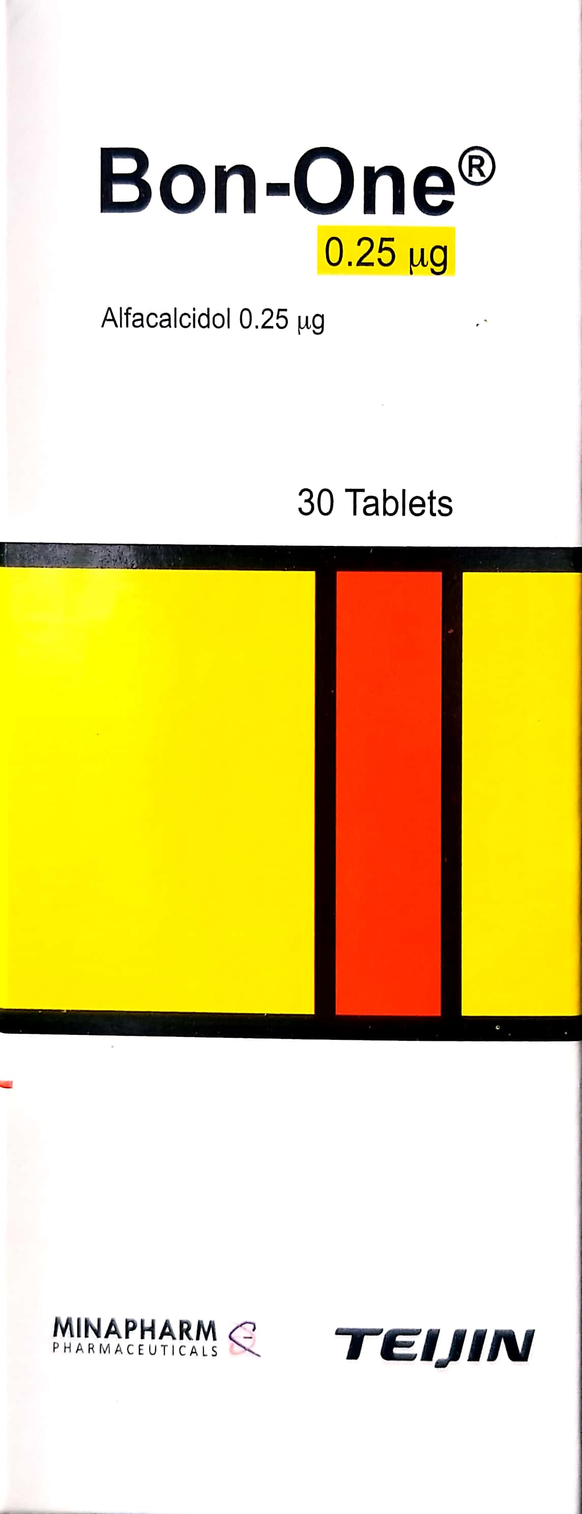
The management of a navicular stress fracture requires a multidisciplinary approach that integrates diagnostic precision, pharmacological support, and specialized orthopedic intervention. To optimize bone healing, clinicians may prescribe Calcium Gluconate / غلوكونات الكالسيوم 10ml and Bon-one / بون-ون 0.25mcg to address metabolic bone health, while ensuring non-weight-bearing status through the use of Axillary (Underarm) Crutches / عكازات إبطية (أدوات ومساعدات الحركة (عكازات/كراسي)) and appropriate protective equipment like the Pediatric Hip Spica Cast Protector / واقي جبيرة الورك الفخذية للأطفال (أدوات ومساعدات الحركة (عكازات/كراسي)). In cases where conservative measures fail and surgical stabilization becomes necessary, the procedure may involve the use of a Battery Powered Orthopedic Drill/Saw System / نظام مثقاب/منشار عظمي يعمل بالبطارية and a Cortical Bone Screw (2.7mm, 3.5mm, 4.5mm) / برغي عظم قشري (2.7 مم، 3.5 مم، 4.5 مم) to achieve internal fixation, a technique distinct from Maxillofacial ORIF (Open Reduction Internal Fixation) / رد مفتوح وتثبيت داخلي للوجه والفكين (ORIF) (عملية كبرى في غرف العمليات) or [Alveolar Bone Grafting / تطعيم العظم السنخي (عملية كبرى في غرف العمليات)](https