Clinical Assessment & Protocol
Typical Presentation (HPI)
EN: Patient presents with a known history of SLE, reporting a flare of polyarthropathy characterized by symmetrical joint pain, morning stiffness lasting >60 minutes, and localized swelling. Symptoms involve [specify joints, e.g., PIP, MCP, wrists], exacerbated by activity and improved with rest. Denies current fever, malar rash, or pleuritic chest pain. AR: يراجع المريض بتاريخ معروف للإصابة بالذئبة الحمامية الجهازية (SLE)، مع شكوى من نوبة التهاب مفاصل متعددة تتميز بألم مفصلي متناظر، وتيبس صباحي يستمر لأكثر من 60 دقيقة، وتورم موضعي. تشمل الأعراض [حدد المفاصل، مثل: المفاصل بين السلاميات القريبة، المفاصل المشطية السلامية، الرسغين]، وتزداد حدة مع النشاط وتتحسن بالراحة. ينفي المريض وجود حمى حالية، أو طفح جلدي فراشي، أو ألم صدري جنبي.
General Examination
EN: General: Patient is alert and oriented, in no acute distress. Musculoskeletal: Symmetrical synovitis noted in [specify joints]. Tenderness to palpation present over [specify joints] with limited range of motion due to pain. No evidence of joint deformity or erosive changes. Skin: No active malar rash, discoid lesions, or photosensitivity noted at this time. AR: الحالة العامة: المريض واعٍ ومدرك للزمان والمكان، ولا يبدو عليه ضيق حاد. الجهاز العضلي الهيكلي: لوحظ وجود التهاب غشاء زليلي متناظر في [حدد المفاصل]. يوجد إيلام عند الجس في [حدد المفاصل] مع محدودية في مدى الحركة بسبب الألم. لا توجد علامات لتشوه المفاصل أو تغيرات تآكلية. الجلد: لا يوجد طفح جلدي فراشي نشط، أو آفات قرصية، أو حساسية ضوئية في الوقت الحالي.
Treatment Protocol
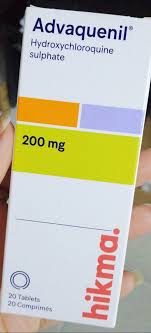
EN: Initiate/Adjust therapy with [Hydroxychloroquine 200-400mg daily / NSAIDs for symptom control / Prednisone taper]. Monitor for potential side effects. Advise strict adherence to medication regimen. Follow up in [timeframe] for repeat labs including CBC, ESR, CRP, and renal function. AR: البدء/تعديل العلاج بـ [هيدروكسي كلوروكين 200-400 ملغ يومياً / مضادات الالتهاب غير الستيرويدية للسيطرة على الأعراض / تقليل تدريجي للبريدنيزون]. مراقبة الآثار الجانبية المحتملة. التأكيد على الالتزام الصارم بنظام الدواء. المتابعة بعد [حدد الفترة الزمنية] لإعادة الفحوصات المخبرية بما في ذلك صورة الدم الكاملة (CBC)، ومعدل ترسب الكريات الحمراء (ESR)، والبروتين التفاعلي (CRP)، ووظائف الكلى.
Patient Education
EN: SLE is a chronic autoimmune condition; polyarthropathy is a common manifestation. Protect skin from UV exposure using high-SPF sunscreen and protective clothing. Report any new symptoms such as fever, severe fatigue, chest pain, or vision changes immediately. Maintain a balanced diet and engage in low-impact physical activity as tolerated. AR: الذئبة الحمامية الجهازية هي حالة مناعية ذاتية مزمنة، ويعد التهاب المفاصل المتعدد أحد أعراضها الشائعة. يجب حماية الجلد من التعرض للأشعة فوق البنفسجية باستخدام واقي شمس ذي معامل حماية عالٍ وملابس واقية. يجب الإبلاغ فوراً عن أي أعراض جديدة مثل الحمى، أو التعب الشديد، أو ألم الصدر، أو تغيرات في الرؤية. حافظ على نظام غذائي متوازن ومارس نشاطاً بدنياً خفيفاً حسب القدرة.
Systemic & Specialized Examinations
EN: Intact distally. AR: سليم طرفياً.
Orthopedic & Trauma Assessments
EN: Idiopathic inflammatory cascade or hematogenous bacterial seeding. AR: سلسلة التهابات مناعية أو انتشار بكتيري عبر الدم.
EN: Non-ambulatory due to severe joint pain on loading. AR: غير قادر على المشي بسبب الألم الشديد عند تحميل الوزن.
EN: Joint is markedly erythematous, warm, tense, and visibly swollen. Loss of skin wrinkles over the joint. AR: المفصل شديد الاحمرار، دافئ، مشدود، ومتورم بوضوح. فقدان تجاعيد الجلد فوق المفصل.
EN: N/A. Clinical picture dominates. AR: لا ينطبق. الصورة السريرية هي السائدة.
EN: Pseudoparalysis. AR: شلل كاذب.
EN: Intact. AR: سليم.
EN: Deferred. AR: مؤجل.
EN: 2+ symmetric. AR: 2+ متماثلة.
Comprehensive Clinical Guide: Systemic Lupus Erythematosus (SLE) with Polyarthropathy
1. Introduction and Overview
Systemic Lupus Erythematosus (SLE) is a prototypical multisystem autoimmune disease characterized by the loss of self-tolerance, leading to the production of autoantibodies against nuclear antigens, the formation of immune complexes, and subsequent end-organ damage. When SLE presents with polyarthropathy—defined as the inflammation of five or more joints—it represents one of the most common and debilitating manifestations of the disease.
Polyarthropathy in SLE is distinct from the erosive, destructive arthritis seen in Rheumatoid Arthritis (RA). It is typically non-erosive, migratory, and symmetric, affecting the small joints of the hands, wrists, and knees. However, a subset of patients may develop Jaccoud’s arthropathy, a deforming condition caused by ligamentous laxity rather than bone erosion. Understanding the interplay between systemic immune dysregulation and musculoskeletal involvement is critical for the orthopedic specialist, rheumatologist, and primary care physician.
2. Deep-Dive: Mechanisms and Pathophysiology
The pathophysiology of SLE-associated polyarthropathy is a complex cascade involving both innate and adaptive immune pathways.
The Immunological Cascade
- Loss of Tolerance: Defective clearance of apoptotic debris results in an accumulation of nuclear antigens (DNA, histones, Ro/La proteins).
- Autoantibody Production: B-cell hyperactivity leads to the production of Anti-Nuclear Antibodies (ANA) and anti-double-stranded DNA (anti-dsDNA) antibodies.
- Immune Complex Deposition: These antibodies form immune complexes that deposit in the synovial lining of joints.
- Complement Activation: The classical complement pathway is activated (C1q, C4, C3), leading to the recruitment of neutrophils and macrophages into the synovial space.
- Cytokine Storm: Locally produced TNF-alpha, IL-1, and IL-6 contribute to synovial hyperplasia and inflammation.
The "Jaccoud’s" Mechanism
Unlike RA, which features pannus formation and cartilage degradation, SLE polyarthropathy often involves chronic inflammation of the periarticular structures (tendons, ligaments, and joint capsules). This leads to:
* Fibrosis: Chronic inflammation induces tissue shortening.
* Laxity: Repeated inflammatory insults weaken the structural integrity of the collateral ligaments.
* Deformity: The resulting "reducible" deformities (ulnar deviation, swan-neck deformities) occur without the radiographic evidence of erosive bone destruction.
3. Clinical Indications, Presentation, and Staging
Standard Clinical Presentation
Patients typically present with "flares" of joint pain that mirror systemic disease activity.
| Feature | Description |
|---|---|
| Symmetry | Bilateral involvement of proximal interphalangeal (PIP) and metacarpophalangeal (MCP) joints. |
| Morning Stiffness | Usually lasts >30 minutes; improves with activity. |
| Migratory Nature | Pain may shift from one joint group to another over days/weeks. |
| Systemic Signs | Concurrent malar rash, photosensitivity, alopecia, or pleuritis. |
Clinical Staging (SLE Disease Activity Index - SLEDAI)
Clinicians utilize the SLEDAI to grade the severity of the polyarthropathy and systemic involvement.
- Mild: Transient arthralgia, minimal synovitis, controlled by hydroxychloroquine and NSAIDs.
- Moderate: Persistent polyarthritis, morning stiffness, requiring low-dose corticosteroids or methotrexate.
- Severe (Refractory): Deforming arthropathy (Jaccoud’s), erosive changes (rare), or systemic vasculitic involvement, requiring biologic therapy (Belimumab, Rituximab).
4. Differential Diagnosis
Distinguishing SLE polyarthropathy from other rheumatic conditions is paramount for therapeutic success.
| Condition | Distinguishing Feature |
|---|---|
| Rheumatoid Arthritis (RA) | Positive RF/ACPA; bone erosions on X-ray; persistent synovitis. |
| Psoriatic Arthritis | Dactylitis ("sausage digits"); nail pitting; distal interphalangeal (DIP) involvement. |
| Viral Arthritis | Acute onset, usually self-limiting (e.g., Parvovirus B19, Hepatitis C). |
| Osteoarthritis | Heberden’s/Bouchard’s nodes; lack of systemic inflammation markers (ESR/CRP). |
5. Diagnostic Protocol: Key Tests
A systematic approach is required to confirm SLE with polyarthropathy.
- Serology:
- ANA: Highly sensitive (>95%).
- Anti-dsDNA/Anti-Smith: Highly specific for SLE.
- Complement levels (C3, C4): Low levels indicate active immune complex consumption.
- Inflammatory Markers: ESR (Erythrocyte Sedimentation Rate) and CRP (C-Reactive Protein). Note: CRP may be disproportionately low in SLE compared to RA unless serositis is present.
- Imaging:
- Radiographs: Used primarily to rule out erosive changes (RA) or fractures.
- Musculoskeletal Ultrasound (MSKUS): The "Gold Standard" for detecting subclinical synovitis, tenosynovitis, and power-doppler flow indicating active inflammation.
- MRI: Reserved for cases of suspected osteonecrosis (often caused by corticosteroid use).
6. Risks, Side Effects, and Contraindications
Managing SLE polyarthropathy requires balancing immunosuppression with long-term safety.
- Corticosteroids (Prednisone): Essential for acute flares but carry risks of osteoporosis, avascular necrosis (AVN), diabetes, and hypertension.
- Hydroxychloroquine (HCQ): The cornerstone therapy. Risk: Retinopathy (requires annual ophthalmologic screening).
- Methotrexate/Azathioprine: Risks include hepatotoxicity and myelosuppression.
- Biologics (Belimumab): Increased risk of serious infections.
- Contraindications: Live vaccines should be avoided in patients on moderate-to-high dose immunosuppressants. NSAIDs should be used with caution in patients with lupus nephritis due to potential renal perfusion impairment.
7. Long-Term Prognosis and Management
The prognosis for SLE polyarthropathy is generally favorable regarding joint function if treated early. Early initiation of Disease-Modifying Antirheumatic Drugs (DMARDs) prevents the progression to Jaccoud’s arthropathy.
- Maintenance: Hydroxychloroquine should be continued indefinitely to prevent flares.
- Monitoring: Regular assessment of renal function (Urinalysis/Protein-Creatinine Ratio) and hematologic profiles.
- Rehabilitation: Physical and occupational therapy are vital to maintain joint range of motion and prevent muscle atrophy.
8. Massive FAQ Section
Q1: Is SLE arthritis permanent?
A: In most cases, it is reversible. However, if chronic inflammation leads to laxity (Jaccoud’s), some joint deformity may persist despite medical control.
Q2: How does SLE arthritis differ from RA?
A: SLE arthritis is typically non-erosive. While RA causes bone destruction, SLE causes ligamentous and capsular changes that lead to deformity.
Q3: Can I take NSAIDs for my lupus joint pain?
A: Yes, but only under physician supervision. Patients with lupus nephritis must avoid NSAIDs as they can impact kidney function.
Q4: What is the role of ultrasound in SLE?
A: Ultrasound is highly sensitive for detecting synovial thickening and increased vascularity (Doppler flow) before clinical symptoms are obvious.
Q5: Why is my CRP normal if my joints hurt?
A: Unlike RA, SLE does not always produce a massive elevation in CRP. A normal CRP does not rule out active SLE.
Q6: Does SLE cause bone death?
A: SLE itself rarely causes bone death, but the long-term use of corticosteroids for SLE treatment is a major risk factor for Avascular Necrosis (AVN).
Q7: Can diet help with SLE polyarthropathy?
A: While no diet cures SLE, an anti-inflammatory diet (rich in Omega-3 fatty acids) may help modulate the systemic inflammatory response.
Q8: When should I see an orthopedic surgeon?
A: Orthopedic consultation is necessary if there is suspicion of AVN, severe joint instability, or if joint function significantly impairs daily living despite medical management.
Q9: Does pregnancy affect SLE arthritis?
A: Pregnancy can trigger flares or stabilize the disease. Women with SLE require high-risk obstetric monitoring to manage medication safety and disease activity.
Q10: Is fatigue related to the joint pain?
A: Yes, fatigue is a core symptom of SLE. It is often multifactorial, caused by systemic inflammation, anemia, or the psychological burden of a chronic diagnosis.
9. Conclusion
Systemic Lupus Erythematosus with polyarthropathy requires a multidisciplinary approach. By combining aggressive serological monitoring, advanced imaging, and a nuanced understanding of immune-mediated versus degenerative joint changes, clinicians can significantly improve patient quality of life. The shift from reactive to proactive management—utilizing early DMARD therapy—remains the gold standard in preventing long-term disability and structural joint failure.