Clinical Assessment & Protocol
Typical Presentation (HPI)
EN: Patient presents for follow-up of fracture nonunion. Reports persistent localized pain, functional impairment, and inability to bear weight/use extremity as expected for duration of injury. Denies recent trauma, fever, or chills. Previous interventions include [ORIF/casting/external fixation] performed on [Date]. Current pain level is [X]/10, exacerbated by movement and loading. AR: يراجع المريض للمتابعة بسبب عدم التئام الكسر. يشكو من ألم موضعي مستمر، وعجز وظيفي، وعدم القدرة على تحمل الوزن أو استخدام الطرف كما هو متوقع بالنسبة لمدة الإصابة. ينفي وجود إصابات حديثة أو حمى أو قشعريرة. تشمل التدخلات السابقة [تثبيت جراحي داخلي/تجبير/تثبيت خارجي] التي أجريت بتاريخ [التاريخ]. مستوى الألم الحالي هو [X]/10، ويزداد مع الحركة والتحميل.
General Examination
EN: Inspection reveals [atrophy/swelling/deformity] at the fracture site. Palpation demonstrates localized tenderness, abnormal mobility at the fracture site, and absence of clinical callus formation. Neurovascular status is intact distally with palpable pulses and normal capillary refill. Range of motion is limited by pain and mechanical instability. AR: يظهر الفحص وجود [ضمور/تورم/تشوه] في موقع الكسر. يظهر الجس وجود إيلام موضعي، وحركة غير طبيعية في موقع الكسر، وغياب تشكل الدشبذ العظمي السريري. الحالة العصبية الوعائية سليمة في الأطراف البعيدة مع نبض محسوس وزمن إعادة ملء شعري طبيعي. مدى الحركة محدود بسبب الألم وعدم الاستقرار الميكانيكي.
Treatment Protocol
EN: Plan: 1. Obtain repeat imaging (X-rays/CT) to assess for biological vs. mechanical nonunion. 2. Discuss revision surgery (bone grafting, hardware exchange, or compression plating). 3. Consider adjunctive therapies (bone stimulator). 4. Maintain activity modifications as directed. 5. Follow up in [X] weeks for review of imaging and surgical planning. AR: الخطة: 1. إجراء تصوير شعاعي (أشعة سينية/أشعة مقطعية) لتقييم ما إذا كان عدم الالتئام بيولوجياً أم ميكانيكياً. 2. مناقشة الجراحة التصحيحية (طعم عظمي، استبدال الأدوات الجراحية، أو التثبيت بالضغط). 3. النظر في العلاجات المساعدة (محفز نمو العظام). 4. الالتزام بتعديلات النشاط حسب التوجيهات. 5. المتابعة بعد [X] أسبوعاً لمراجعة التصوير والتخطيط الجراحي.
Patient Education
EN: Nonunion occurs when a fracture fails to heal within the expected timeframe. This may be due to biological factors (blood supply, nutrition, smoking) or mechanical factors (instability at the fracture site). It is critical to avoid weight-bearing or strenuous activity unless cleared. Smoking cessation is mandatory to improve bone healing potential. Report any signs of infection, such as increasing redness, warmth, or drainage. AR: يحدث عدم الالتئام عندما يفشل الكسر في الالتئام خلال الإطار الزمني المتوقع. قد يعود ذلك إلى عوامل بيولوجية (تروية دموية، تغذية، تدخين) أو عوامل ميكانيكية (عدم استقرار في موقع الكسر). من الضروري تجنب تحمل الوزن أو النشاط الشاق ما لم يُسمح بذلك. الإقلاع عن التدخين إلزامي لتحسين فرص التئام العظام. يجب الإبلاغ عن أي علامات للعدوى، مثل زيادة الاحمرار، أو الحرارة، أو وجود إفرازات.
Systemic & Specialized Examinations
EN: Crucial evaluation: Median, Ulnar, and Radial nerves INTACT to light touch and 2-point discrimination. AIN/PIN/Radial motor functions normal. AR: تقييم حاسم: العصب الأوسط، الزندي، والكعبري سليمة. الوظائف الحركية للأعصاب سليمة.
Orthopedic & Trauma Assessments
EN: FOOSH injury or high-impact direct trauma. AR: إصابة السقوط على يد ممدودة أو صدمة مباشرة عالية التأثير.
EN: Normal. Ambulatory. AR: طبيعية.
EN: Marked soft tissue swelling and ecchymosis over the fracture site. Characteristic 'Dinner Fork' deformity (if distal radius) or gross angulation. AR: تورم وكدمات ملحوظة فوق موقع الكسر. تشوه 'شوكة العشاء' المميز (إذا كان في الكعبرة) أو تقوس إجمالي.
EN: N/A for acute fracture. AR: لا ينطبق للكسر الحاد.
EN: Hand intrinsic and extrinsic tendons function properly. AR: أوتار اليد الداخلية والخارجية تعمل بشكل صحيح.
EN: Sensation 100% intact globally. AR: الإحساس سليم 100%.
EN: Deferred. AR: مؤجل.
EN: Radial and Ulnar pulses strong (2+). Capillary refill brisk (< 2 sec). AR: النبض الكعبري والزندي قوي. عودة امتلاء الشعيرات سريعة.
Comprehensive Clinical Guide: Nonunion of Fracture
1. Introduction and Clinical Overview
Nonunion of a fracture represents a critical failure in the orthopedic healing process, occurring when the natural osteogenic potential of the bone is insufficient to bridge the fracture gap. In clinical terms, a fracture is defined as a nonunion when it has failed to show progressive signs of healing on serial radiographs for a period of at least six to nine months post-injury.
Unlike delayed union—where healing is simply slower than expected—nonunion implies that the biological repair process has reached a standstill. Without surgical or biological intervention, the fracture is unlikely to heal spontaneously. This condition poses significant challenges to patient mobility, quality of life, and healthcare resource utilization, often requiring complex revision surgeries and prolonged rehabilitation.
2. Etiology and Pathophysiology
The pathophysiology of nonunion is multifactorial, involving a complex interplay between mechanical stability, biological environment, and systemic patient factors.
The Diamond Concept of Fracture Healing
To understand nonunion, one must reference the "Diamond Concept," which posits that bone healing requires four pillars:
1. Osteoconduction: A scaffold for bone growth.
2. Osteoinduction: Growth factors to stimulate progenitor cells.
3. Osteogenesis: Viable bone-forming cells.
4. Mechanical Stability: A stable environment for callus formation.
Primary Etiological Categories
- Mechanical Factors: Excessive motion at the fracture site (instability) prevents the formation of a stable callus, leading to hypertrophic nonunion.
- Biological Factors: Compromised vascularity, soft tissue damage, infection, or systemic metabolic deficiencies lead to atrophic nonunion.
- Patient-Related Factors: Tobacco use, diabetes mellitus, malnutrition, vitamin D deficiency, and the use of NSAIDs or corticosteroids.
3. Clinical Staging and Classification
The Weber and Cech classification system is the gold standard for categorizing nonunions based on biological activity, as visualized on radiographic imaging.
| Type | Classification | Characteristics | Biological Potential |
|---|---|---|---|
| Hypertrophic | "Elephant Foot" | Excessive callus formation; gap is bridged by fibrocartilage. | High (Good blood supply) |
| Oligotrophic | Non-reactive | Minimal callus formation; ends are rounded and sclerotic. | Moderate |
| Atrophic | "Pencil Point" | Resorption of bone ends; lack of biological activity. | Very Low (Poor blood supply) |
- Hypertrophic Nonunion: Typically caused by mechanical instability. The "Elephant Foot" appearance is indicative of the bone attempting to heal but failing due to micromotion.
- Atrophic Nonunion: Represents a biological failure. The bone ends become necrotic or resorbed, often secondary to severe trauma, infection, or compromised soft tissue.
4. Standard Clinical Presentation
Patients presenting with a nonunion often report symptoms that deviate from the expected recovery trajectory. Key clinical markers include:
- Persistent Pain: Chronic, localized pain that worsens with weight-bearing or movement.
- Clinical Instability: Palpable motion at the fracture site (pseudarthrosis).
- Functional Limitation: Inability to return to pre-injury activities of daily living.
- Deformity: Visible or palpable angulation or shortening at the fracture site.
- Soft Tissue Issues: Recurrent swelling, skin tethering, or persistent drainage (if infection is present).
5. Diagnostic Methodology
A systematic approach is required to differentiate nonunion from delayed union and to identify the underlying cause.
Key Diagnostic Tests
- Radiographic Imaging (X-rays): Serial AP and lateral views are mandatory. Look for persistence of the fracture line, lack of bridging callus, and sclerosis of the fracture ends.
- Computed Tomography (CT): Highly sensitive for assessing bridging callus in complex areas (e.g., scaphoid, tibial plateau) where plain films are obscured.
- Laboratory Analysis: Inflammatory markers (ESR, CRP) are critical to rule out subclinical osteomyelitis.
- MRI: Useful for evaluating soft tissue health and vascularity, though metal artifacts from previous hardware can limit utility.
- Nuclear Medicine (Bone Scans): Used to assess metabolic activity, though less specific than CT/MRI.
6. Differential Diagnosis
It is essential to distinguish nonunion from other clinical entities:
* Delayed Union: The fracture is healing, but at a rate slower than the clinical average.
* Malunion: The fracture has healed, but in an anatomically unacceptable position (angulation, rotation, or shortening).
* Osteomyelitis: Infection of the bone, which may mimic atrophic nonunion or cause secondary nonunion.
* Pathological Fracture: Fracture occurring through bone weakened by tumor or metabolic bone disease (e.g., Paget’s disease).
7. Management and Therapeutic Approaches
Management is dictated by the biological classification of the nonunion.
- Hypertrophic Management: The primary goal is mechanical stability. This often involves compression plating or exchange nailing to eliminate motion at the fracture site.
- Atrophic Management: The primary goal is biological enhancement. This requires debridement of necrotic bone, stabilization, and the addition of bone graft (autograft, allograft, or bone morphogenetic proteins/BMPs).
- Infected Nonunion: Requires a two-stage approach: debridement and antibiotic-impregnated cement spacers, followed by definitive fixation once the infection is eradicated.
8. Risks and Contraindications
Intervening in a nonunion carries significant risks:
* Hardware Failure: Revision surgery increases the risk of screw loosening or plate breakage.
* Infection: Repeated surgical entries increase the risk of iatrogenic osteomyelitis.
* Donor Site Morbidity: If harvesting an autograft (e.g., iliac crest), patients may experience chronic donor site pain or nerve injury.
* Contraindications: Surgery may be contraindicated in patients with severe peripheral vascular disease, uncontrolled systemic infection, or limited physiological reserve where the risk of mortality outweighs the benefit of surgery.
9. Long-term Prognosis
The prognosis for nonunion is generally favorable if the underlying biological and mechanical triggers are addressed. However, patients must be informed that:
* Revision surgery is often more complex than the primary procedure.
* Recovery times are significantly longer.
* The risk of chronic pain or permanent functional deficit remains, especially in high-energy fractures or cases involving extensive bone loss.
10. Frequently Asked Questions (FAQ)
1. What is the difference between delayed union and nonunion?
Delayed union is a fracture that takes longer than average to heal but is still progressing. Nonunion is a complete cessation of the healing process where no further biological progress is observed.
2. Can smoking cause a nonunion?
Yes. Nicotine is a potent vasoconstrictor that reduces oxygen delivery to the fracture site, severely inhibiting the bone’s ability to generate new tissue.
3. What is the "Elephant Foot" nonunion?
This is a term used for hypertrophic nonunion. It refers to the radiographic appearance of abundant callus that fails to bridge the gap due to excessive motion.
4. How is infection ruled out in a nonunion case?
Clinicians use blood work (CRP/ESR levels) and, if necessary, a bone biopsy or aspiration to culture the site for bacteria.
5. Is bone grafting always necessary?
Not always. In hypertrophic nonunion, stabilizing the bone is often sufficient. In atrophic nonunion, grafting is usually required to provide the biological components (osteoconduction/induction) necessary for healing.
6. What role does Vitamin D play in fracture healing?
Vitamin D is essential for calcium absorption and bone mineralization. Deficiency is a common systemic factor that delays or prevents bone healing.
7. Can NSAIDs prevent a fracture from healing?
Some studies suggest that prolonged use of high-dose NSAIDs (like ibuprofen or naproxen) can inhibit prostaglandin production, which is a key part of the bone healing cascade.
8. How does diabetes affect nonunion risk?
Diabetes impairs microvascular circulation and alters the inflammatory response, both of which are critical for the initial stages of callus formation.
9. What is an "Exchange Nailing"?
This is a technique where a previously placed intramedullary nail is removed and replaced with a larger diameter nail to increase stability and stimulate healing.
10. What is the success rate of treating a nonunion?
Success rates vary widely based on the location of the bone and the cause of the nonunion, but with appropriate mechanical stabilization and biological grafting, success rates typically exceed 80–90%.
11. Clinical Conclusion
Nonunion of a fracture is a complex orthopedic complication that demands a rigorous diagnostic workup and a tailored treatment plan. By identifying whether the failure is mechanical (hypertrophic) or biological (atrophic), the orthopedic surgeon can implement targeted interventions. Patient education regarding lifestyle modifications—specifically smoking cessation and glycemic control—is as vital as the surgical procedure itself. Through a combination of precise fixation and biological augmentation, the majority of nonunions can be successfully resolved, restoring function and quality of life to the patient.
Related Clinical Integration
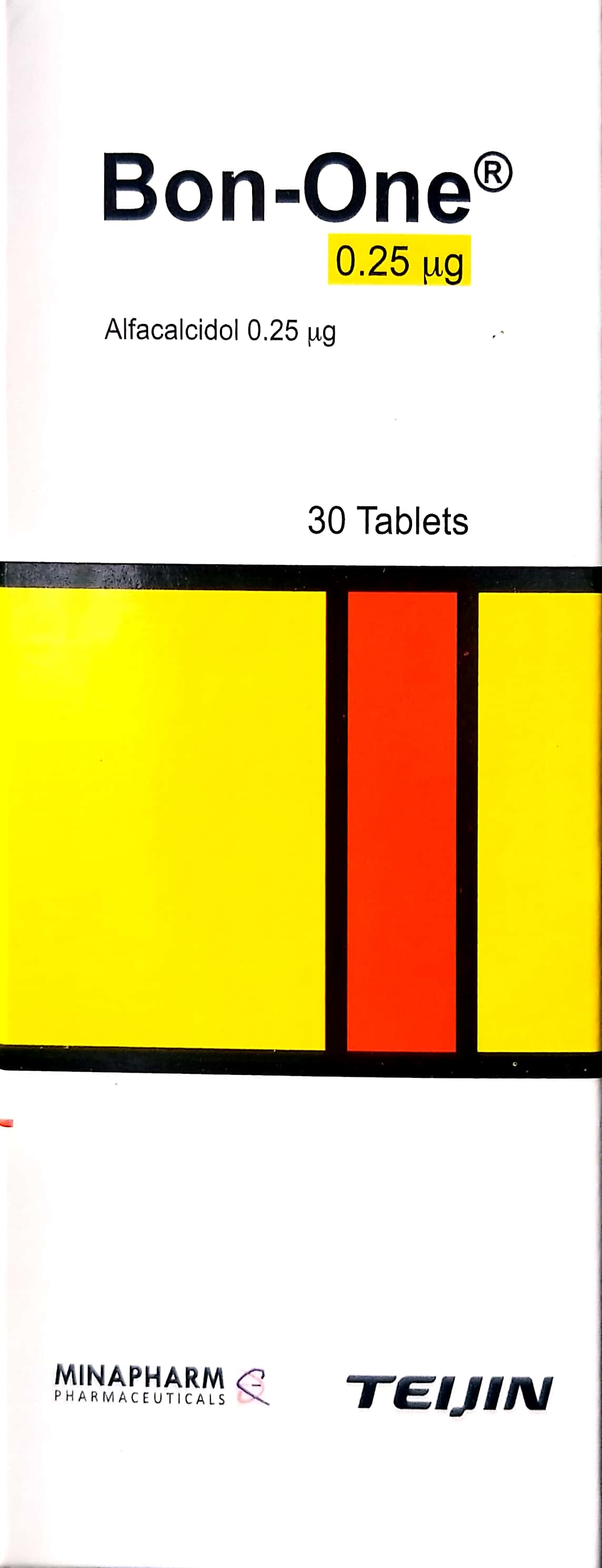
In the modern clinical management of nonunion of fractures, a multidisciplinary approach is essential to restore biological and mechanical stability, often requiring a combination of pharmacological support, advanced surgical techniques, and specialized instrumentation. To address the underlying metabolic deficiencies that impede healing, clinicians may prescribe osteoinductive or bone-building agents such as Bon-one / بون-ون 0.25mcg or Forteo / فورتيو 20mcg/dose. When surgical intervention is indicated, the use of precision tools like the Flexible Osteotome System / نظام مبضع عظمي مرن facilitates the preparation of the nonunion site for biological augmentation, which may include procedures such as Alveolar Bone Grafting / تطعيم العظم السنخي (عملية كبرى في غرف العمليات). For practitioners seeking deeper insights into evidence-based protocols, we recommend reviewing our comprehensive resources on Delayed Union and Nonunion of Fractures: Comprehensive Surgical Management, Operative Management of Fracture Nonunions: A Comprehensive Guide, Advanced Management of Fracture Nonunions: Biomechanics, Bone Grafting, and Surgical Reconstruction, Management of Fracture Nonunions: A Surgical Guide, Nonunion Fractures: Orthopedic Epidemiology, Biomechanics, & Classification, and the specialized study on [اندماج مفصل إبهام القدم بتطعيم العظام: دليل شامل لاستعادة وظيفة القدم مع الأستاذ الدكتور محمد هطيف](https://www.hutaifortho.com/ar/hub/%D8%A