Clinical Assessment & Protocol
Typical Presentation (HPI)
EN: Patient presents for routine follow-up of senile osteoporosis. Denies acute bone pain, recent falls, or symptoms suggestive of pathological fracture. No history of height loss or kyphotic deformity progression. Adherent to current calcium and vitamin D supplementation. AR: يراجع المريض للمتابعة الدورية لهشاشة العظام المرتبطة بالسن. ينفي وجود آلام عظمية حادة، أو حوادث سقوط حديثة، أو أعراض تشير إلى وجود كسر مرضي. لا يوجد تاريخ لفقدان الطول أو تطور في تشوه تقوس الظهر. المريض ملتزم بتناول مكملات الكالسيوم وفيتامين د الحالية.
General Examination
EN: General appearance: Alert and oriented. Musculoskeletal: Gait is steady and non-antalgic. Spine: No midline tenderness, no evidence of acute vertebral collapse or new kyphosis. Extremities: Full range of motion without focal tenderness. Neurological: Intact sensation and motor strength, no signs of cord compression. AR: المظهر العام: واعٍ ومدرك للزمان والمكان. الجهاز العضلي الهيكلي: المشية متزنة ولا يوجد عرج. العمود الفقري: لا يوجد ألم عند الجس في خط المنتصف، ولا توجد علامات لانهيار فقاري حاد أو تقوس جديد. الأطراف: مدى حركي كامل بدون نقاط ألم موضعية. الجهاز العصبي: الإحساس والقوة العضلية سليمان، لا توجد علامات لانضغاط الحبل الشوكي.
Treatment Protocol
EN: Continue daily supplementation of Calcium (1200mg) and Vitamin D3 (800-2000 IU). Initiate/Continue anti-resorptive therapy (e.g., Bisphosphonates) as per bone mineral density (BMD) trends. Schedule annual DEXA scan to monitor T-score progression. Fall prevention counseling provided. AR: الاستمرار في تناول مكملات الكالسيوم (1200 ملغ) وفيتامين د3 (800-2000 وحدة دولية) يومياً. البدء/الاستمرار في العلاج المضاد لارتشاف العظم (مثل البايفوسفونيت) وفقاً لنتائج قياس كثافة العظام (BMD). جدولة فحص كثافة العظام (DEXA) سنوياً لمراقبة تطور درجة T-score. تم تقديم إرشادات حول الوقاية من السقوط.
Patient Education
EN: Focus on weight-bearing exercises to maintain bone density. Ensure adequate dietary intake of calcium-rich foods. Implement home safety modifications to prevent falls (e.g., removing rugs, improving lighting). Report any new severe back pain or sudden mobility limitations immediately. AR: التركيز على تمارين تحمل الوزن للحفاظ على كثافة العظام. ضمان تناول كميات كافية من الأطعمة الغنية بالكالسيوم. إجراء تعديلات السلامة المنزلية لمنع السقوط (مثل إزالة السجاد، وتحسين الإضاءة). الإبلاغ فوراً عن أي ألم شديد جديد في الظهر أو محدودية مفاجئة في الحركة.
Systemic & Specialized Examinations
EN: Crucial evaluation: Median, Ulnar, and Radial nerves INTACT to light touch and 2-point discrimination. AIN/PIN/Radial motor functions normal. AR: تقييم حاسم: العصب الأوسط، الزندي، والكعبري سليمة. الوظائف الحركية للأعصاب سليمة.
Orthopedic & Trauma Assessments
EN: FOOSH injury or high-impact direct trauma. AR: إصابة السقوط على يد ممدودة أو صدمة مباشرة عالية التأثير.
EN: Normal. Ambulatory. AR: طبيعية.
EN: Marked soft tissue swelling and ecchymosis over the fracture site. Characteristic 'Dinner Fork' deformity (if distal radius) or gross angulation. AR: تورم وكدمات ملحوظة فوق موقع الكسر. تشوه 'شوكة العشاء' المميز (إذا كان في الكعبرة) أو تقوس إجمالي.
EN: N/A for acute fracture. AR: لا ينطبق للكسر الحاد.
EN: Hand intrinsic and extrinsic tendons function properly. AR: أوتار اليد الداخلية والخارجية تعمل بشكل صحيح.
EN: Sensation 100% intact globally. AR: الإحساس سليم 100%.
EN: Deferred. AR: مؤجل.
EN: Radial and Ulnar pulses strong (2+). Capillary refill brisk (< 2 sec). AR: النبض الكعبري والزندي قوي. عودة امتلاء الشعيرات سريعة.
Clinical Guide: Senile Osteoporosis (ICD-10 M81.0)
1. Comprehensive Introduction & Overview
Senile osteoporosis, clinically classified under ICD-10 as M81.0 (Osteoporosis without current pathological fracture), represents a systemic skeletal disorder characterized by low bone mass and microarchitectural deterioration of bone tissue. Unlike postmenopausal osteoporosis, which is primarily driven by estrogen deficiency, senile osteoporosis typically manifests in patients aged 70 and older. It is a multi-factorial condition where the rate of bone resorption exceeds the rate of bone formation, leading to increased skeletal fragility.
The absence of a "current pathological fracture" is a critical diagnostic distinction. It suggests that while the patient’s bone mineral density (BMD) has crossed the threshold for clinical concern (T-score ≤ -2.5), the structural integrity has not yet failed under physiological load. This stage represents a vital window of opportunity for clinical intervention, lifestyle modification, and pharmacological management to prevent the inevitable morbidity associated with fragility fractures.
2. Deep-Dive: Etiology and Pathophysiology
The pathophysiology of senile osteoporosis is rooted in the "age-related decline" of homeostatic mechanisms. It is not merely the result of one hormone deficiency but a confluence of cellular and systemic changes.
The Cellular Mechanism
- Osteoblast Senescence: As individuals age, mesenchymal stem cells in the bone marrow show a decreased capacity to differentiate into osteoblasts. This leads to a reduction in bone formation.
- Osteoclast Hyperactivity: While osteoblast activity wanes, osteoclast activity remains constant or increases due to secondary hyperparathyroidism, leading to a net loss of bone.
- RANK/RANKL/OPG Pathway: The imbalance between RANKL (which promotes osteoclastogenesis) and OPG (which inhibits it) is exacerbated by chronic low-grade inflammation, often termed "inflammaging."
Systemic Contributors
| Factor | Mechanism of Action |
|---|---|
| Vitamin D Deficiency | Reduced intestinal calcium absorption leading to secondary hyperparathyroidism. |
| Calcium Malabsorption | Age-related decline in gut efficiency and dietary intake. |
| Hormonal Changes | Decline in IGF-1, growth hormone, and adrenal androgens. |
| Physical Inactivity | Lack of mechanical loading (Wolff’s Law) leads to disuse atrophy. |
3. Clinical Staging and Diagnostic Criteria
Diagnosis is primarily established through Dual-Energy X-ray Absorptiometry (DXA) scanning. The World Health Organization (WHO) provides the standard classification for T-scores.
Table: WHO T-Score Classification
| Classification | T-Score Definition |
|---|---|
| Normal | -1.0 or higher |
| Osteopenia | Between -1.0 and -2.5 |
| Osteoporosis | -2.5 or lower |
| Severe Osteoporosis | -2.5 or lower + history of fragility fracture |
Note: In the context of M81.0, the patient meets the -2.5 threshold but is excluded from the "Severe" category due to the lack of a current fracture.
4. Standard Presentation and Clinical Assessment
Patients with senile osteoporosis are often asymptomatic until a fracture occurs. However, clinical suspicion should be high in elderly patients presenting with:
* Loss of Height: A loss of >2cm since young adulthood or >1cm in a single year.
* Kyphosis: Progressive thoracic curvature (Dowager’s hump).
* Back Pain: Often chronic, non-specific axial pain that may indicate micro-fractures of the vertebral bodies.
* Functional Decline: Reduced grip strength and balance issues.
Laboratory Evaluation
To rule out secondary causes (e.g., hyperthyroidism, multiple myeloma, or renal osteodystrophy), the following labs are standard:
* Complete Blood Count (CBC)
* Comprehensive Metabolic Panel (Calcium, Phosphorus, Albumin)
* 25-hydroxyvitamin D levels
* Thyroid Stimulating Hormone (TSH)
* Serum Protein Electrophoresis (if myeloma is suspected)
5. Differential Diagnosis
Distinguishing senile osteoporosis from other skeletal pathologies is imperative:
1. Osteomalacia: Often due to severe Vitamin D deficiency; characterized by "soft" bones rather than low density.
2. Multiple Myeloma: Presents with lytic lesions and significant bone pain; requires screening via serum protein electrophoresis.
3. Hyperparathyroidism: Can mimic osteoporosis but is characterized by elevated serum calcium and PTH.
4. Metastatic Bone Disease: Should be considered if the patient has a history of malignancy.
6. Management and Long-Term Prognosis
The goal is to maintain bone density and improve fall prevention.
Pharmacological Management
- Bisphosphonates (e.g., Alendronate, Risedronate): First-line therapy to inhibit osteoclast-mediated bone resorption.
- RANK Ligand Inhibitors (e.g., Denosumab): Highly effective in increasing BMD by preventing osteoclast maturation.
- Anabolic Agents (e.g., Teriparatide, Romosozumab): Reserved for patients at very high risk, as they stimulate new bone formation.
Lifestyle and Safety
- Calcium & Vitamin D: Supplementation is mandatory if dietary intake is insufficient.
- Weight-Bearing Exercise: Resistance training to stimulate osteoblastic activity.
- Fall Prevention: Home modifications (removing rugs, improving lighting) and balance training (Tai Chi, physical therapy).
7. Risks, Side Effects, and Contraindications
While pharmacological intervention is vital, clinicians must be aware of potential complications:
- Bisphosphonates: Risk of Osteonecrosis of the Jaw (ONJ) and atypical femoral fractures (long-term use). Patients should maintain strict oral hygiene.
- Denosumab: Risk of rebound hypercalcemia upon discontinuation. It must be followed by bisphosphonate therapy if stopped.
- Anabolic Agents: Contraindicated in patients with a history of radiation therapy to the skeleton or Paget’s disease.
8. Massive FAQ Section
Q1: Does a T-score of -2.5 automatically mean I will break a bone?
A: No. It indicates that your bone density is in the osteoporotic range, which increases your risk, but it is not a guarantee of a fracture. Proper management significantly lowers this risk.
Q2: Can I reverse senile osteoporosis?
A: While you can rarely return to "normal" bone density, you can significantly improve bone quality and density, which dramatically reduces fracture risk.
Q3: How often should I have a DXA scan?
A: Typically, every 1–2 years depending on your treatment plan and the severity of the bone loss.
Q4: Do I need to stop taking my medication if I feel fine?
A: Absolutely not. Osteoporosis is a "silent" disease. The medication is working to keep your bones strong, even if you feel no physical changes.
Q5: Is dairy the only way to get calcium?
A: No. Leafy greens (kale, collards), fortified plant milks, sardines, and almonds are excellent sources. Supplements can be used if dietary goals aren't met.
Q6: What is the role of Vitamin D in bone health?
A: Vitamin D is essential for the absorption of calcium in the gut. Without it, your body cannot use the calcium you consume, leading to weak bones.
Q7: Can exercise make my osteoporosis worse?
A: Only if you engage in high-impact activities or spinal flexion (bending forward) without professional supervision. Low-impact, weight-bearing exercise is highly encouraged.
Q8: What is an "atypical femoral fracture"?
A: It is a rare fracture that can occur in the thigh bone after long-term bisphosphonate use. It is why we periodically reassess the need for continued treatment (a "drug holiday").
Q9: Does smoking affect my bone density?
A: Yes. Nicotine and other toxins in cigarettes are toxic to bone cells and interfere with the absorption of calcium.
Q10: Are there any natural supplements that cure osteoporosis?
A: No. While supplements like K2 or Magnesium can support bone health, they are not a substitute for FDA-approved pharmacological treatment in diagnosed osteoporosis.
9. Clinical Conclusion
Senile osteoporosis (M81.0) is a manageable chronic condition. The primary objective for the clinician is to identify the patient early, quantify the risk, and implement a multimodal strategy involving pharmacological stability, nutritional optimization, and aggressive fall prevention. By addressing the physiological decline associated with aging, we can transition the patient from a state of fragility to a state of skeletal resilience, thereby preventing the transition from M81.0 to a fracture-related diagnosis.
Disclaimer: This guide is for educational purposes for healthcare professionals and clinical students. It does not replace institutional protocols or individualized clinical judgment. Always consult current guidelines from the American Association of Clinical Endocrinologists (AACE) or the Endocrine Society for the most recent updates on pharmacotherapy.
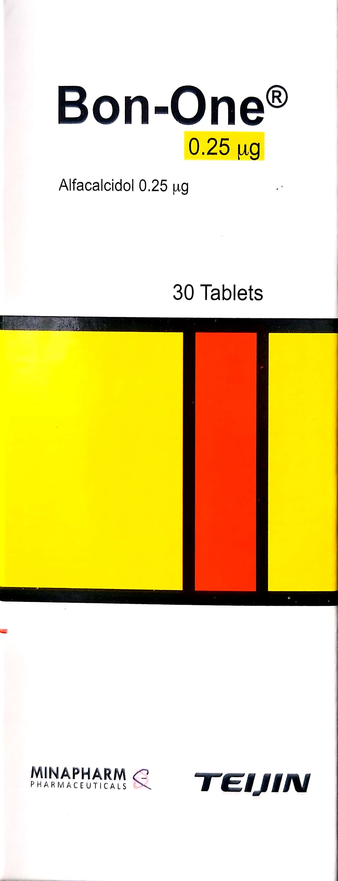
Related Clinical Integration
In the management of senile osteoporosis without current pathological fracture, the clinical objective shifts toward proactive bone density optimization and fall prevention to mitigate future morbidity. Pharmacological intervention is central to this strategy, utilizing agents such as Bon-one / بون-ون 0.25mcg for calcium homeostasis and Prolia / بروليا 60 mg/mL to inhibit osteoclast-mediated bone resorption. For patients exhibiting gait instability or those at high risk of secondary injury, the use of a CAM Walker Boot (Walking Boot) / حذاء المشي الطبي (حذاء ووكر) (أدوات ومساعدات الحركة (عكازات/كراسي)) provides necessary mechanical support to prevent acute trauma. Clinicians should further integrate these interventions with a robust understanding of bone health, as detailed in our resources on Osteoporosis in Orthopedics: Biomechanics, Fracture Patterns & Surgical Management and [الوقاية من هشاشة العظام تبدأ مبكرًا: دليل شامل لصحة عظام أطفالك](https://www.hutaifortho.com/ar/hub/%D8%A7%D9%84%D8%AA%D8%B5%D8%AF%D9%8A-%D9%84%D9%85%D8%B4%D9%83%D9%84%D8%A9-%D9%87%D8%B4%D8%A7%D8%B4%D8%A9-%D8%A7%D9%84%D8%B9%D8%B8%D8%A7%D9%85-%D8%A7%D9%84%D9%85%D8%AA%D9%86%D8%A7%D9%85%D9%8A%D8%A9-%D8%AF%D9%84%D9%8A%D9%84-%D8%B4%D8%A7%D9%85%D9%84-%