Clinical Assessment & Protocol
Typical Presentation (HPI)
EN: Patient presents with chronic, progressive left hip pain, localized to the groin and lateral hip, exacerbated by weight-bearing activities and prolonged ambulation. Reports morning stiffness lasting <30 minutes, relieved by rest. Denies trauma, fevers, or radicular symptoms. Pain interferes with activities of daily living (ADLs) and sleep. AR: يعاني المريض من ألم مزمن ومتفاقم في الورك الأيسر، يتركز في منطقة الأربية والجانب الخارجي للورك، ويزداد سوءاً مع الأنشطة التي تتطلب تحمل الوزن والمشي لفترات طويلة. يشكو من تيبس صباحي يستمر لأقل من 30 دقيقة ويتحسن بالراحة. لا توجد إصابات سابقة أو حمى أو أعراض عصبية جذرية. الألم يؤثر على الأنشطة اليومية والنوم.
General Examination
EN: Left hip exam: Antalgic gait noted. Tenderness to palpation over the left greater trochanter and groin. Range of motion (ROM) is restricted in internal rotation and abduction, with crepitus noted on passive manipulation. Positive FADIR and FABER tests. Neurovascular status intact distally. AR: فحص الورك الأيسر: لوحظ وجود مشية متألمة. يوجد ألم عند الجس فوق المدور الكبير الأيسر ومنطقة الأربية. مدى الحركة محدود في الدوران الداخلي والإبعاد، مع وجود فرقعة عند التحريك السلبي. اختبارات FADIR و FABER إيجابية. الحالة العصبية الوعائية سليمة في الأطراف البعيدة.
Treatment Protocol
EN: Conservative management initiated: Activity modification, physical therapy for hip abductor strengthening, and weight management. Pharmacotherapy includes scheduled NSAIDs or acetaminophen as tolerated. Consider intra-articular corticosteroid or hyaluronic acid injection if symptoms persist. Discussed surgical consultation for total hip arthroplasty (THA) if conservative measures fail. AR: تم البدء بالعلاج التحفظي: تعديل الأنشطة، العلاج الطبيعي لتقوية عضلات الورك المبعدة، وإدارة الوزن. يشمل العلاج الدوائي مضادات الالتهاب غير الستيرويدية أو الباراسيتامول حسب التحمل. يُنظر في حقن الكورتيكوستيرويد أو حمض الهيالورونيك داخل المفصل إذا استمرت الأعراض. تمت مناقشة الاستشارة الجراحية لعملية استبدال مفصل الورك الكلي في حال فشل الإجراءات التحفظية.
Patient Education
EN: Primary osteoarthritis is a degenerative condition of the hip joint. Focus on low-impact exercises (swimming, cycling) to maintain joint mobility. Avoid high-impact activities. Use assistive devices (cane) if necessary to offload the joint. Monitor for worsening pain, night pain, or functional decline, and report to clinic immediately. AR: الفصال العظمي الأولي هو حالة تنكسية في مفصل الورك. ركز على التمارين منخفضة التأثير (السباحة، ركوب الدراجات) للحفاظ على مرونة المفصل. تجنب الأنشطة عالية التأثير. استخدم أدوات مساعدة (عكاز) إذا لزم الأمر لتخفيف الحمل عن المفصل. راقب أي زيادة في الألم، أو ألم ليلي، أو تراجع في القدرة الوظيفية، وراجع العيادة فوراً في حال حدوث ذلك.
Systemic & Specialized Examinations
EN: Intact distally. AR: سليم طرفياً.
Orthopedic & Trauma Assessments
EN: Degenerative joint disease. No acute trauma. AR: تآكل تنكسي في المفصل. لا توجد صدمة.
EN: Antalgic/Trendelenburg gait. Reduced stance phase on the affected side. AR: مشية متألمة/ترندلينبورغ. قصر مرحلة الوقوف على الجانب المصاب.
EN: Gluteal/quadriceps atrophy may be present chronologically. AR: قد يوجد ضمور في الأرداف/الرباعية مع الوقت.
EN: FABER: POSITIVE (groin pain). FADIR: POSITIVE. Thomas Test: Positive for flexion contracture. AR: اختبارات فابر وفادير: إيجابية (ألم في المغبن). اختبار توماس: إيجابي لانكماش الانثناء.
EN: Weak hip abductors (positive Trendelenburg). AR: ضعف في مبعدات الورك (ترندلينبورغ إيجابي).
EN: Intact. AR: سليم.
EN: 2+ symmetric. AR: 2+ متماثلة.
EN: Distal pulses 2+. AR: النبضات الطرفية طبيعية.
Comprehensive Clinical Guide: Primary Osteoarthritis of the Left Hip (Coxarthrosis)
1. Introduction and Overview
Primary Osteoarthritis (OA) of the left hip, clinically referred to as idiopathic coxarthrosis, represents a degenerative, non-inflammatory joint disease characterized by the progressive erosion of articular cartilage, subchondral bone remodeling, and osteophyte formation. Unlike secondary osteoarthritis, which arises from identifiable trauma, congenital dysplasia, or metabolic disorders, primary OA is considered an age-related, multifactorial process involving genetic predisposition, mechanical wear, and low-grade systemic inflammation.
The hip joint, a classic ball-and-socket synovial joint, is uniquely susceptible to the degenerative forces of weight-bearing. When the protective cartilage of the left acetabulum and femoral head degrades, the resulting bone-on-bone friction leads to significant morbidity, characterized by chronic pain, functional limitation, and a substantial decrease in the patient’s quality of life.
2. Technical Specifications and Pathophysiology
The Mechanics of Cartilage Degeneration
The pathophysiology of primary hip OA is a complex biological cascade rather than simple "wear and tear." The process begins at the molecular level within the chondrocyte, the sole cell type responsible for maintaining the extracellular matrix (ECM) of articular cartilage.
- ECM Degradation: The imbalance between anabolic (repair) and catabolic (degradation) processes leads to the upregulation of matrix metalloproteinases (MMPs) and aggrecanases. These enzymes break down the collagen type II network and proteoglycans.
- Subchondral Bone Remodeling: As cartilage thins, mechanical load is redistributed to the underlying subchondral bone. This triggers osteoblast activity, resulting in subchondral sclerosis (increased bone density) and the formation of subchondral cysts (geodes).
- Osteophyte Formation: The periosteum at the joint margins responds to increased mechanical stress by forming bone spurs (osteophytes), which attempt to stabilize the joint but ultimately restrict range of motion (ROM) and cause impingement.
Clinical Staging: The Kellgren-Lawrence (K-L) System
The K-L scale remains the gold standard for grading the radiographic severity of hip OA:
| Grade | Description | Clinical Correlation |
|---|---|---|
| 0 | Normal | No radiographic features of OA. |
| 1 | Doubtful | Possible osteophyte lipping, joint space narrowing questionable. |
| 2 | Mild | Definite osteophytes, possible joint space narrowing. |
| 3 | Moderate | Multiple osteophytes, definite joint space narrowing, some sclerosis. |
| 4 | Severe | Large osteophytes, marked joint space narrowing, severe sclerosis, bone deformity. |
3. Clinical Indications and Presentation
Standard Presentation
Patients presenting with primary OA of the left hip typically report a insidious onset of symptoms. The clinical "classic" presentation includes:
- Pain Localization: Typically felt in the groin, lateral hip, or referred pain to the anterior thigh or knee. Pain radiating to the knee is a common clinical trap that can lead to misdiagnosis.
- Stiffness: Morning stiffness lasting less than 30 minutes, or stiffness following periods of inactivity (the "gel phenomenon").
- Functional Decline: Difficulty with activities of daily living (ADLs) such as putting on socks, clipping toenails, or rising from a low chair.
- Gait Abnormalities: A Trendelenburg gait or an antalgic gait (shortened stance phase on the left side) due to pain-induced avoidance.
Diagnostic Workup
A definitive diagnosis requires a combination of physical examination and imaging.
- Physical Exam (The "C-Sign"): The patient cups their hand around the lateral hip (the C-sign), indicating deep hip pain.
- Provocative Tests:
- FADIR Test (Flexion, Adduction, Internal Rotation): Highly sensitive for intra-articular pathology.
- FABER Test (Flexion, Abduction, External Rotation): Often reproduces pain in the hip or sacroiliac joint.
- Imaging:
- Anteroposterior (AP) Pelvis: Essential for comparing the left hip to the asymptomatic right side.
- Lateral/Frog-leg view: Useful for assessing the femoral head-neck junction.
- MRI: Generally not required for standard OA diagnosis, but useful for ruling out labral tears or avascular necrosis (AVN).
4. Differential Diagnosis
It is critical to distinguish primary OA from other pathologies that mimic hip pain:
- Trochanteric Bursitis: Pain is localized to the greater trochanter; usually tender to palpation, unlike intra-articular OA.
- Lumbar Radiculopathy (L3-L4): Pain radiates to the hip/thigh but is associated with dermatomal sensory changes or reflex deficits.
- Femoroacetabular Impingement (FAI): Often seen in younger, athletic populations; involves anatomical morphology (cam/pincer) that leads to secondary OA.
- Avascular Necrosis (AVN): Characterized by rapid progression and specific risk factors (steroid use, alcohol consumption, sickle cell).
5. Risks, Contraindications, and Prognosis
Therapeutic Risks and Contraindications
Management strategies carry inherent risks:
* NSAIDs: Long-term use presents risks of gastrointestinal bleeding, renal impairment, and cardiovascular events.
* Intra-articular Corticosteroids: Risk of localized infection (septic arthritis), transient hyperglycemia in diabetics, and potential cartilage toxicity with repeated injections.
* Surgical Intervention (Total Hip Arthroplasty): Risks include deep vein thrombosis (DVT), pulmonary embolism, prosthetic dislocation, periprosthetic fracture, and infection.
Long-Term Prognosis
Primary OA is a progressive condition. While it is rarely life-threatening, the prognosis for mobility is variable. Early intervention—focusing on weight management, physical therapy (PT), and activity modification—can significantly delay the need for surgical intervention. For patients with end-stage (K-L Grade 4) disease, Total Hip Arthroplasty (THA) is the gold standard, offering a high success rate (over 90% at 15-20 years) in restoring function and eliminating pain.
6. FAQ: Frequently Asked Questions
1. Is primary osteoarthritis of the left hip hereditary?
Yes, genetics play a significant role. Studies suggest that hereditary factors account for approximately 50-60% of the risk for developing hip OA.
2. Can physical therapy cure my hip osteoarthritis?
PT cannot "cure" cartilage loss, but it is highly effective at managing symptoms, improving joint stability, and delaying the progression of functional disability by strengthening the peri-articular musculature.
3. Why do I feel pain in my knee if the problem is in my hip?
This is known as "referred pain." The hip and knee share similar nerve pathways (specifically the obturator and femoral nerves). The brain often misinterprets signals from the hip joint as originating from the knee.
4. When is the right time to consider surgery?
Surgery is considered when non-operative management (PT, NSAIDs, weight loss) fails to provide sufficient relief to maintain a reasonable quality of life or when ADLs are severely restricted.
5. Are dietary supplements like Glucosamine/Chondroitin effective?
Clinical evidence is mixed. While some patients report subjective improvement, major clinical trials (such as the GAIT study) have shown results comparable to placebos for moderate-to-severe OA.
6. Will walking make my hip OA worse?
Moderate, low-impact exercise is encouraged. However, high-impact activities (running, jumping) on hard surfaces can accelerate symptoms. Cycling and swimming are generally recommended.
7. Does the weather affect my hip pain?
Many patients report increased pain during changes in barometric pressure. While not fully understood, it is believed that changes in pressure affect the pressure within the synovial fluid of the joint.
8. What is the difference between primary and secondary OA?
Primary OA is idiopathic and age-related. Secondary OA is caused by a specific pre-existing condition, such as hip dysplasia, a previous fracture, or metabolic diseases like hemochromatosis.
9. How long is the recovery after a total hip replacement?
Most patients are up and walking with assistance on the day of surgery. Full recovery, including the return to normal activities and strengthening, typically takes 3 to 6 months.
10. Can I prevent hip OA from getting worse?
While you cannot stop the biological clock, maintaining a healthy BMI is the single most effective way to reduce the load on the hip joint, thereby slowing the rate of cartilage degradation.
7. Conclusion
Primary osteoarthritis of the left hip is a manageable condition if addressed with a multi-modal approach. As an expert in the field, it is clear that patient education is the cornerstone of successful management. By understanding the mechanical and biological drivers of the disease, patients can make informed decisions regarding lifestyle modifications, pharmacological intervention, and, when necessary, surgical consultation. The goal is not merely to "treat the X-ray," but to optimize the patient’s functional capacity and overall health.
Related Clinical Integration
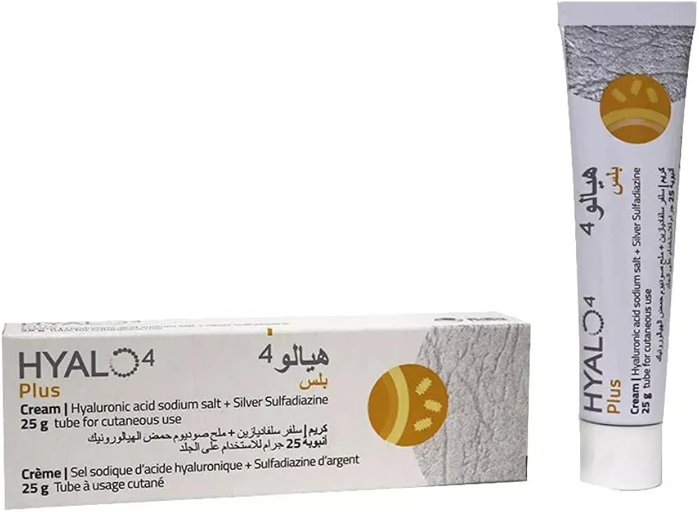
In the management of Primary Osteoarthritis of the left hip, a comprehensive clinical approach integrates pharmacological, surgical, and rehabilitative strategies to optimize patient outcomes. Initial conservative management often involves the administration of anti-inflammatory agents such as Celcox / سيلكوكس 100mg or Meloxicam / ميلوكسيكام 25mg, while Hyalo4 plus cream / هيالو 4 بلس كريم 0.2% / 1% may be utilized for adjunctive wound care. When conservative measures fail to mitigate structural degradation, surgical intervention becomes necessary, utilizing specialized tools like the Oscillating Bone Saw Blade (Wide, Narrow, Deep Cut) to perform procedures such as Hip Resurfacing Arthroplasty / رأب مفصل الورك بالترميم السطحي (عملية كبرى في غرف العمليات) or Total Hip Arthroplasty (THA) / استبدال مفصل الورك الكلي (THA) (عملية كبرى في غرف العمليات). Post-operative recovery and mobility are supported through the use of assistive devices, including Axillary (Underarm) Crutches / عكازات إبطية (أدوات ومساعدات الحركة (عكازات/كراسي)) and the CAM Walker Boot (Walking Boot) / حذاء المشي الطبي (حذاء ووكر) (أدوات ومساعدات الحركة (عكازات/كراسي)). For further clinical insight, practitioners and patients are encouraged to review our educational resources, including the