Clinical Assessment & Protocol
Typical Presentation (HPI)
EN: Patient presents with chronic, progressive right hip pain, localized to the groin and lateral hip, exacerbated by weight-bearing activities and prolonged standing. Reports morning stiffness lasting <30 minutes, improved with mild activity. Denies trauma, fevers, or radicular symptoms. Pain interferes with activities of daily living and sleep. AR: يعاني المريض من ألم مزمن ومتفاقم في مفصل الورك الأيمن، يتركز في منطقة الأربية والجانب الخارجي للورك، ويزداد سوءاً مع الأنشطة التي تتطلب تحمل الوزن والوقوف لفترات طويلة. يشكو من تيبس صباحي يستمر لأقل من 30 دقيقة، ويتحسن مع الحركة الخفيفة. لا يوجد تاريخ لإصابات، أو حمى، أو أعراض عصبية جذرية. الألم يؤثر على الأنشطة اليومية والنوم.
General Examination
EN: Right hip examination reveals antalgic gait. Palpation demonstrates tenderness over the greater trochanter and groin. Range of motion (ROM) is restricted in internal rotation and abduction, with crepitus noted on passive manipulation. FABER and FADIR tests are positive for pain. Neurovascular status is intact distally. AR: فحص الورك الأيمن يظهر مشية متألمة. يظهر الجس وجود ألم عند الضغط على المدور الكبير ومنطقة الأربية. مدى الحركة محدود في الدوران الداخلي والإبعاد، مع وجود فرقعة عند التحريك السلبي. اختبارات FABER و FADIR إيجابية للألم. الحالة العصبية الوعائية سليمة في الأطراف البعيدة.
Treatment Protocol
EN: Conservative management initiated: weight-bearing modification, physical therapy for abductor strengthening and ROM maintenance, and non-steroidal anti-inflammatory drugs (NSAIDs) as needed. Discussed potential for intra-articular corticosteroid or viscosupplementation injections if refractory to conservative measures. AR: تم البدء بالعلاج التحفظي: تعديل الأنشطة التي تتطلب تحمل الوزن، العلاج الطبيعي لتقوية العضلات المبعدة والحفاظ على مدى الحركة، ومضادات الالتهاب غير الستيرويدية (NSAIDs) عند الحاجة. تمت مناقشة إمكانية الحقن الموضعي بالكورتيكوستيرويد أو المكملات اللزوجية داخل المفصل في حال عدم الاستجابة للعلاجات التحفظية.
Patient Education
EN: Primary osteoarthritis is a degenerative condition of the hip joint cartilage. Focus on low-impact exercise (swimming, cycling) to maintain joint mobility. Maintain healthy weight to reduce mechanical load on the hip. Monitor for worsening pain, night pain, or functional decline, which may necessitate surgical consultation (total hip arthroplasty). AR: الفصال العظمي الأولي هو حالة تنكسية لغضروف مفصل الورك. يُنصح بالتركيز على التمارين منخفضة التأثير (السباحة، ركوب الدراجات) للحفاظ على مرونة المفصل. الحفاظ على وزن صحي لتقليل الحمل الميكانيكي على الورك. يجب مراقبة أي تفاقم في الألم، أو ألم ليلي، أو تراجع في الوظيفة الحركية، مما قد يستدعي استشارة جراحية (لإجراء عملية استبدال مفصل الورك).
Systemic & Specialized Examinations
EN: Intact distally. AR: سليم طرفياً.
Orthopedic & Trauma Assessments
EN: Degenerative joint disease. No acute trauma. AR: تآكل تنكسي في المفصل. لا توجد صدمة.
EN: Antalgic/Trendelenburg gait. Reduced stance phase on the affected side. AR: مشية متألمة/ترندلينبورغ. قصر مرحلة الوقوف على الجانب المصاب.
EN: Gluteal/quadriceps atrophy may be present chronologically. AR: قد يوجد ضمور في الأرداف/الرباعية مع الوقت.
EN: FABER: POSITIVE (groin pain). FADIR: POSITIVE. Thomas Test: Positive for flexion contracture. AR: اختبارات فابر وفادير: إيجابية (ألم في المغبن). اختبار توماس: إيجابي لانكماش الانثناء.
EN: Weak hip abductors (positive Trendelenburg). AR: ضعف في مبعدات الورك (ترندلينبورغ إيجابي).
EN: Intact. AR: سليم.
EN: 2+ symmetric. AR: 2+ متماثلة.
EN: Distal pulses 2+. AR: النبضات الطرفية طبيعية.
Comprehensive Clinical Guide: Primary Osteoarthritis of the Right Hip
1. Introduction and Overview
Primary Osteoarthritis (OA) of the hip, clinically classified under ICD-10 code M16.11, represents a chronic, progressive, degenerative joint disease characterized by the mechanical failure of the articular cartilage within the right coxofemoral joint. Unlike secondary osteoarthritis, which is precipitated by identifiable trauma, congenital dysplasia, or systemic metabolic disease, primary (idiopathic) osteoarthritis is considered a multifactorial process involving age-related senescence, genetic predisposition, and cumulative biomechanical stress.
The hip joint is a ball-and-socket synovial joint, and when the hyaline cartilage—which serves as the primary load-bearing surface—undergoes biochemical and structural degradation, the underlying subchondral bone undergoes compensatory remodeling. This guide provides an exhaustive clinical overview of the pathophysiology, diagnostic pathways, and management strategies for this condition.
2. Etiology and Pathophysiology
The degradation of the hip joint in primary OA is not merely a "wear-and-tear" phenomenon; it is a complex metabolic event involving the entire joint organ.
The Pathophysiological Cascade:
- Chondrocyte Activation: The initial insult involves the activation of chondrocytes, which shift from an anabolic state (synthesizing Type II collagen and proteoglycans) to a catabolic state.
- Enzymatic Degradation: Elevated levels of matrix metalloproteinases (MMPs) and aggrecanases (ADAMTS-5) begin to cleave the extracellular matrix (ECM).
- Subchondral Bone Remodeling: As cartilage thins, the subchondral bone experiences increased mechanical pressure. This leads to microfractures, vascular invasion, and the formation of osteophytes (bone spurs) at the joint margins.
- Synovial Inflammation: Low-grade chronic synovitis often develops, where the synovial membrane releases pro-inflammatory cytokines (IL-1, TNF-alpha), further accelerating cartilage degradation.
Risk Factors:
| Risk Factor Type | Specific Variables |
|---|---|
| Non-Modifiable | Age (>50), Female Gender, Genetic Predisposition (Family history) |
| Modifiable | Obesity (BMI >30), High-impact repetitive loading, Metabolic Syndrome |
| Biomechanical | Leg length discrepancy, Femoral neck-shaft angle variations |
3. Clinical Staging and Grading
The clinical severity of right hip OA is typically categorized using the Kellgren-Lawrence (K-L) Grading Scale, which relies on radiographic findings.
| Grade | Description | Radiographic Findings |
|---|---|---|
| 0 | None | Normal joint space, no osteophytes. |
| 1 | Doubtful | Possible osteophytic lipping, questionable joint space narrowing. |
| 2 | Mild | Definite osteophytes, possible joint space narrowing. |
| 3 | Moderate | Moderate joint space narrowing, multiple osteophytes, bony sclerosis. |
| 4 | Severe | Marked joint space narrowing, severe sclerosis, deformity of bone ends. |
4. Standard Clinical Presentation
Patients presenting with primary OA of the right hip typically report a predictable symptom cluster:
- Pain Distribution: Classically reported in the groin area, but often radiates to the anterior thigh or the knee (referred pain).
- Morning Stiffness: Generally lasts less than 30 minutes.
- Mechanical Symptoms: "Giving way" or catching sensations during gait.
- Functional Limitations: Difficulty with activities of daily living (ADLs) such as putting on socks, tying shoelaces, or entering/exiting a vehicle.
- Gait Abnormalities: Development of an antalgic gait or a Trendelenburg gait (due to abductor weakness).
5. Differential Diagnosis
It is imperative to distinguish primary hip OA from other pathologies that mimic hip pain:
- Lumbar Radiculopathy (L3-L4): Often presents with referred pain to the hip; ruled out via neurological exam and MRI of the lumbar spine.
- Trochanteric Bursitis: Pain is lateral and point-tender; hip joint range of motion (ROM) is typically preserved.
- Avascular Necrosis (AVN): Often presents in younger patients with specific risk factors (steroid use, alcohol); appears distinct on MRI (crescent sign).
- Femoroacetabular Impingement (FAI): Often precedes OA; characterized by CAM or PINCER morphology.
6. Diagnostic Testing
A multi-modal approach is required for a definitive diagnosis:
- Physical Examination:
- ROM Assessment: Internal rotation is almost always the first movement to be lost.
- FABER Test: (Flexion, Abduction, External Rotation) – pain indicates hip pathology.
- Scour Test: Provocative test for intra-articular hip pain.
- Imaging:
- Anteroposterior (AP) Pelvis Radiograph: The gold standard for assessing joint space narrowing and osteophyte formation.
- Lateral/Frog-leg View: Essential to visualize the femoral head-neck junction.
- MRI: Reserved for cases where physical exam is inconsistent with X-ray findings or to rule out labral tears/AVN.
7. Long-Term Prognosis and Management
Primary OA is a progressive condition. While there is no "cure" that reverses cartilage loss, long-term management focuses on functional preservation.
Conservative Management (The First Line):
- Physical Therapy: Strengthening the gluteus medius, quadriceps, and core musculature to reduce joint load.
- Weight Management: Reducing BMI to decrease compressive forces on the acetabulum.
- Pharmacotherapy: NSAIDs (e.g., Naproxen, Ibuprofen) for acute flares; acetaminophen for baseline pain management.
- Assistive Devices: Utilizing a cane in the contralateral (left) hand to reduce the joint reaction force on the right hip.
Surgical Intervention (Total Hip Arthroplasty - THA):
When conservative measures fail and quality of life is severely compromised, THA is the definitive intervention. Modern implants (ceramic-on-polyethylene or ceramic-on-ceramic) have a lifespan exceeding 20-25 years in the majority of patients.
8. Risks, Side Effects, and Contraindications
Risks of Conservative Treatment:
- NSAIDs: Gastrointestinal bleeding, renal impairment, and cardiovascular risk.
- Injections (Corticosteroids): Risk of septic arthritis (rare) or transient hyperglycemia in diabetics.
Risks of Surgical Treatment (THA):
- Infection: Deep prosthetic joint infection (DPI) remains a catastrophic complication.
- DVT/PE: Prophylaxis is mandatory post-operatively.
- Dislocation: Particularly in the first 6 weeks post-op.
- Leg Length Discrepancy: A common patient-reported dissatisfaction factor.
9. Frequently Asked Questions (FAQ)
Q1: Can I reverse hip osteoarthritis through diet?
A: No. While anti-inflammatory diets may reduce systemic inflammation, they cannot regenerate lost hyaline cartilage.
Q2: Is my right hip pain actually a back problem?
A: It is possible. Referred pain from the lumbar spine is common. A physical therapist or orthopedist can perform a "hip-spine syndrome" evaluation to differentiate.
Q3: How much weight should I lose to help my hip?
A: Every pound of weight lost reduces the compressive force across the hip joint by 3-4 pounds during gait. Even a 5-10% body weight reduction can significantly improve symptoms.
Q4: Do I need an MRI for my hip pain?
A: Usually, no. An AP Pelvis X-ray is sufficient for diagnosing primary OA. MRI is typically reserved for surgical planning or identifying soft tissue pathology.
Q5: What is the "Gold Standard" for hip OA pain relief?
A: Total Hip Arthroplasty (THA) is the most effective intervention for end-stage (Grade 4) osteoarthritis.
Q6: Can physical therapy make my pain worse?
A: It is common to experience muscle soreness when starting a new strengthening program, but it should not cause sharp, joint-deep pain. If it does, intensity should be adjusted.
Q7: Will my left hip develop OA too?
A: Because primary OA is often systemic and genetic, there is a high probability of contralateral involvement over time, though it may progress at a different rate.
Q8: Are injections better than surgery?
A: Injections (Corticosteroids or Hyaluronic Acid) are "bridge" therapies meant to manage symptoms, not correct the mechanical defect. Surgery is a permanent mechanical fix.
Q9: How long is the recovery after a hip replacement?
A: Most patients walk the same day as surgery. Full recovery, including tissue healing and gait normalization, typically takes 3-6 months.
Q10: Is swimming good for my right hip?
A: Yes. Aquatic therapy is ideal because it provides resistance for muscle strengthening while the buoyancy of the water eliminates the impact forces on the joint.
10. Clinical Conclusion
Primary Osteoarthritis of the right hip is a debilitating but manageable condition. The key to successful clinical outcomes lies in early diagnosis, a structured physical therapy regimen, and precise timing for surgical intervention when conservative measures no longer provide adequate pain relief or functional mobility. Clinicians should prioritize a "patient-centered" approach, balancing radiographic severity with the patient's individual functional goals.
DISCLAIMER: This document is for educational and informational purposes only and does not constitute medical advice, diagnosis, or treatment. Always seek the advice of your physician or other qualified health provider with any questions you may have regarding a medical condition.
Related Clinical Integration
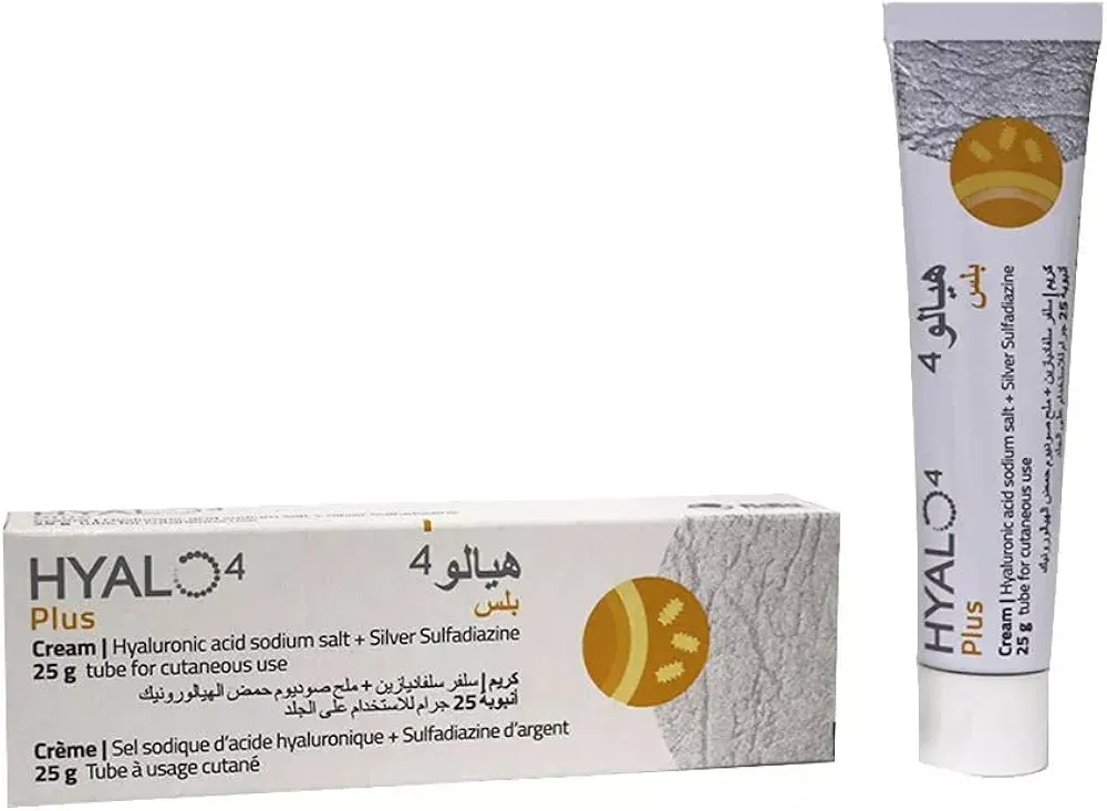
In the management of Primary Osteoarthritis, Right Hip, a multidisciplinary clinical approach is essential to address both symptomatic relief and functional restoration. Initial conservative management often involves pharmacological intervention with anti-inflammatory agents such as Celcox / سيلكوكس 100mg or Meloxicam / ميلوكسيكام 25mg, while Hyalo4 plus cream / هيالو 4 بلس كريم 0.2% / 1% may be utilized for adjunctive wound care or dermatological support. When conservative measures fail to mitigate pain or restore mobility, surgical intervention becomes necessary, utilizing specialized tools like the Oscillating Bone Saw Blade (Wide, Narrow, Deep Cut) to perform procedures such as Hip Resurfacing Arthroplasty / رأب مفصل الورك بالترميم السطحي (عملية كبرى في غرف العمليات) or Total Hip Arthroplasty (THA) / استبدال مفصل الورك الكلي (THA) (عملية كبرى في غرف العمليات). Post-operative recovery and rehabilitation are supported by mobility aids including Axillary (Underarm) Crutches / عكازات إبطية (أدوات ومساعدات الحركة (عكازات/كراسي)) and CAM Walker Boot (Walking Boot) / حذاء المشي الطبي (حذاء ووكر) (أدوات ومساعدات الحركة (عكازات/كراسي)), while clinical decision-making is reinforced by evidence-based resources such as the