Clinical Assessment & Protocol
Typical Presentation (HPI)
EN: Patient presents with acute onset of localized lumbar back pain following [mechanism of injury/minor trauma]. Pain is exacerbated by movement, weight-bearing, and spinal flexion. No reported radiculopathy, bowel/bladder dysfunction, or saddle anesthesia. Pain intensity rated at [X]/10. AR: يراجع المريض بسبب ألم حاد وموضعي في أسفل الظهر عقب [آلية الإصابة/رض خفيف]. يزداد الألم مع الحركة، تحميل الوزن، وثني العمود الفقري. لا توجد شكاوى من اعتلال جذري، خلل في وظائف الأمعاء أو المثانة، أو خدر في منطقة السرج. شدة الألم تقدر بـ [X]/10.
General Examination
EN: Physical examination reveals localized tenderness over the [L1-L5] spinous process. Paraspinal muscle spasm noted. Neurological examination: motor strength 5/5 in bilateral lower extremities, intact sensation to light touch in all dermatomes, deep tendon reflexes 2+ and symmetric. No midline step-off or gross deformity. Gait is guarded but stable. AR: يكشف الفحص السريري عن إيلام موضعي فوق النتوء الشوكي للفقرة [L1-L5]. لوحظ وجود تشنج في العضلات المجاورة للعمود الفقري. الفحص العصبي: القوة العضلية 5/5 في الطرفين السفليين، الإحساس سليم للمس الخفيف في جميع القطاعات الجلدية، المنعكسات الوترية العميقة 2+ ومتناظرة. لا يوجد انزياح في الخط الناصف أو تشوه ظاهر. المشية حذرة ولكنها مستقرة.
Treatment Protocol
EN: Conservative management initiated: activity modification, avoidance of heavy lifting, and bracing as indicated. Analgesic regimen includes NSAIDs and/or acetaminophen for pain control. Physical therapy referral for core stabilization and progressive mobilization. Follow-up imaging in [X] weeks to assess healing. AR: تم البدء بالعلاج التحفظي: تعديل الأنشطة، تجنب رفع الأثقال، واستخدام دعامة الظهر حسب الحاجة. يتضمن نظام المسكنات مضادات الالتهاب غير الستيرويدية و/أو الباراسيتامول للسيطرة على الألم. إحالة للعلاج الطبيعي لتقوية العضلات الجذعية والتحريك التدريجي. إجراء تصوير متابعة بعد [X] أسابيع لتقييم الالتئام.
Patient Education
EN: You have been diagnosed with a stable lumbar compression fracture. Focus on pain management and gradual return to activity. Avoid lifting objects heavier than [X] lbs and avoid twisting or bending at the waist. Seek immediate medical attention if you experience new numbness, weakness in legs, or loss of bowel/bladder control. AR: تم تشخيص إصابتك بكسر انضغاطي مستقر في الفقرات القطنية. ركز على إدارة الألم والعودة التدريجية للنشاط. تجنب رفع أشياء أثقل من [X] رطل، وتجنب الالتواء أو الانحناء عند الخصر. اطلب العناية الطبية الفورية إذا شعرت بخدر جديد، ضعف في الساقين، أو فقدان السيطرة على الأمعاء أو المثانة.
Systemic & Specialized Examinations
EN: Distinct radiculopathy (L4/L5/S1). Strict Cauda Equina precautions documented. AR: اعتلال عصبي جذري واضح. تم توثيق تحذيرات متلازمة ذيل الفرس بصرامة.
Orthopedic & Trauma Assessments
EN: Heavy lifting incident with spinal rotation, or insidious degenerative disc disease. AR: حادث رفع أوزان ثقيلة مع دوران للعمود الفقري، أو انزلاق غضروفي تدريجي.
EN: Antalgic gait. Exhibits a 'list' (sciatic scoliosis) away from the affected side. Difficulty with heel/toe walk. AR: مشية متألمة. يظهر ميلاً (جنف وركي) لتخفيف الضغط. صعوبة في المشي على الكعب/الأصابع.
EN: Loss of normal lumbar lordosis. Severe paraspinal muscle spasm. AR: فقدان التقوس القطني الطبيعي. تشنج عضلي شديد حول الفقرات.
EN: Straight Leg Raise (SLR): Strongly positive at 30-45°. Slump test positive. AR: اختبار رفع الساق المستقيمة (SLR): إيجابي بقوة عند 30-45 درجة.
EN: Weakness (4/5) in EHL (L5) or Plantarflexion (S1). AR: ضعف (4/5) في باسطة الإبهام (L5) أو الثني الأخمصي (S1).
EN: Hypoesthesia to pinprick over the foot dorsum (L5) or lateral border (S1). AR: نقص الإحساس للوخز على ظهر القدم (L5) أو الجانب الوحشي (S1).
EN: Achilles (S1) diminished 1+. Patellar (L4) 2+. AR: منعكس وتر أخيل ضعيف 1+. منعكس الرضفة طبيعي 2+.
EN: DP and PT pulses 2+ symmetric. AR: النبضات الطرفية طبيعية.
Comprehensive Clinical Guide: Lumbar Stable Spinal Compression Fracture
1. Introduction & Overview
A Lumbar Stable Spinal Compression Fracture (VCF) represents a significant clinical entity characterized by the loss of vertebral body height, typically resulting from axial loading and flexion. In the orthopedic and neurosurgical landscape, a "stable" classification is paramount; it implies that the posterior ligamentous complex (PLC) remains intact, and the fracture does not pose an immediate risk of neurological deficit or progressive deformity.
While often associated with geriatric populations and metabolic bone disease, these fractures are increasingly recognized in younger patients following high-energy trauma. The lumbar spine, specifically the thoracolumbar junction (T11-L2), is the most common site of these injuries due to the transition between the rigid thoracic kyphosis and the mobile lumbar lordosis.
2. Etiology and Pathophysiology
Etiology
The etiology of stable lumbar compression fractures is bifurcated into two primary categories:
- Osteoporotic/Metabolic: The most common cause. Decreased bone mineral density (BMD) leads to structural failure under physiologic loads (e.g., sitting down too hard, lifting a light object).
- Traumatic: High-energy mechanisms, such as motor vehicle accidents (MVAs), falls from heights, or athletic injuries, can cause fractures even in healthy bone.
Pathophysiology
The vertebral body is a cancellous bone structure contained within a cortical shell.
1. Axial Loading: Force is transmitted through the anterior and middle columns.
2. Failure of Trabeculae: When the force exceeds the yield strength of the cancellous bone, the trabeculae collapse.
3. Wedge Deformity: The anterior aspect of the vertebral body loses height while the posterior wall remains intact, creating the classic "wedge" morphology.
4. Stability Assessment: Stability is determined by the Denis Three-Column Model. A stable fracture involves only the anterior column. If the middle column is involved, the fracture is considered potentially unstable (Burst Fracture).
| Column | Components | Clinical Relevance in Stable VCF |
|---|---|---|
| Anterior | Anterior longitudinal ligament, anterior 2/3 of vertebral body | Primary site of collapse |
| Middle | Posterior 1/3 of vertebral body, posterior longitudinal ligament | Must remain intact for stability |
| Posterior | Facet joints, pedicles, laminae, ligamentum flavum | Must remain intact for stability |
3. Clinical Presentation and Diagnosis
Standard Presentation
Patients typically present with localized, sharp, midline back pain. The pain is often exacerbated by movement, weight-bearing, or changes in position.
* Physical Exam: Point tenderness over the spinous process of the affected vertebra. Patients may demonstrate a "guarded" gait or reduced range of motion.
* Neurological Status: In a stable fracture, the neurological examination should be normal. Any evidence of radiculopathy, bowel/bladder dysfunction, or lower extremity weakness necessitates an immediate upgrade to "unstable" and urgent surgical consultation.
Diagnostic Modalities
| Test | Purpose | Findings |
|---|---|---|
| Plain Radiographs (AP/Lateral) | Initial Screening | Loss of vertebral body height, kyphotic angulation |
| MRI (T1/T2/STIR) | Gold Standard | Edema (acute vs. chronic), PLC integrity |
| CT Scan | Bone Detail | Assesses comminution, retropulsion, and pedicle integrity |
| DEXA Scan | Metabolic baseline | Assesses underlying osteoporosis severity |
4. Clinical Staging and Classification (AO Spine/TLICS)
The Thoracolumbar Injury Classification and Severity (TLICS) score is the clinical standard. A stable compression fracture generally scores low (Total score ≤ 3).
- Compression (A1/A2): 1 point.
- Neurological Status: Intact (0 points).
- Posterior Ligamentous Complex: Intact (0 points).
5. Management Strategies
Conservative Management
For stable fractures, the primary goal is pain management and early mobilization.
1. Analgesia: Acetaminophen, NSAIDs, or short-term calcitonin (for osteoporotic patients).
2. Bracing: Thoracolumbar Orthosis (TLSO) for comfort, though evidence suggests limited efficacy in preventing long-term deformity.
3. Physical Therapy: Focused on core stabilization, posture correction, and fall prevention.
Minimally Invasive Procedures
If pain persists despite 4–6 weeks of conservative therapy:
* Vertebroplasty: Injection of bone cement (PMMA) into the vertebral body to stabilize the fracture and provide immediate pain relief.
* Kyphoplasty: Use of an inflatable balloon to restore vertebral height before cement injection.
6. Risks, Side Effects, and Contraindications
Risks of Non-Intervention
- Kyphotic Deformity: Progressive loss of height leading to "humpback" appearance.
- Chronic Pain: Persistent pain due to pseudoarthrosis or altered biomechanics.
- Adjacent Level Fractures: Increased loading on vertebrae above and below the fractured level.
Contraindications for Cement Augmentation
- Active Infection: Osteomyelitis or systemic sepsis.
- Coagulopathy: Uncorrected bleeding disorders.
- Posterior Wall Defect: High risk of cement leakage into the spinal canal.
7. Long-Term Prognosis
The prognosis for a stable lumbar compression fracture is generally excellent. Most patients achieve significant pain reduction within 8–12 weeks. However, patients with underlying osteoporosis require aggressive management (bisphosphonates, Teriparatide, Calcium/Vitamin D) to prevent secondary fractures, which carry a significantly higher mortality risk in elderly cohorts.
8. Frequently Asked Questions (FAQ)
Q1: How do I know if my fracture is truly "stable"?
A: Stability is determined by a radiologist or surgeon using CT/MRI. If the posterior column and middle column are intact, the fracture is stable.
Q2: Will I need surgery?
A: Most stable fractures are treated non-surgically. Surgery is reserved for intractable pain or failed conservative management.
Q3: How long does the pain last?
A: Acute pain usually subsides in 6–8 weeks. Residual soreness may persist for 3–6 months.
Q4: Should I wear a back brace?
A: Bracing is used for comfort. It does not "heal" the bone faster, but it helps with pain by limiting motion.
Q5: Can I exercise with a spinal fracture?
A: Gentle walking is encouraged. Avoid heavy lifting, twisting, or high-impact activities until cleared by your physician.
Q6: What is the risk of a secondary fracture?
A: Patients with osteoporosis have a significantly higher risk of subsequent fractures. Compliance with metabolic bone medication is essential.
Q7: Is Vertebroplasty dangerous?
A: When performed by experienced interventionalists, it is highly safe. The primary risk is cement leakage, though this is rare in stable compression fractures.
Q8: Why does my doctor want a DEXA scan?
A: To determine if the fracture was caused by low bone mass. Treating the underlying osteoporosis prevents future injuries.
Q9: Can a compression fracture cause paralysis?
A: In a stable fracture, the risk of paralysis is negligible. If you experience numbness or weakness, seek emergency care immediately.
Q10: What is the "humpback" effect?
A: This is kyphosis. It occurs when the front of the vertebra collapses, causing the spine to curve forward. It can be managed with physical therapy and proper postural habits.
9. Clinical Summary Table: Management Phases
| Phase | Duration | Focus |
|---|---|---|
| Acute | 0–2 weeks | Pain control, rest, bracing, activity modification |
| Sub-acute | 2–8 weeks | Gradual increase in activity, core strengthening |
| Chronic | 8+ weeks | Long-term metabolic bone management, fall prevention |
10. Conclusion
A stable lumbar spinal compression fracture is a manageable condition provided that the diagnosis is accurate and the patient adheres to a structured recovery plan. By focusing on pain mitigation, biomechanical support, and aggressive metabolic management of bone density, clinicians can ensure patients return to their baseline functional status. Always consult with a board-certified spine specialist to ensure your specific fracture morphology aligns with the "stable" classification before proceeding with conservative care.
Disclaimer: This guide is for educational purposes only. It does not constitute medical advice, diagnosis, or treatment. Always seek the advice of your physician or other qualified health provider with any questions regarding a medical condition.
Related Clinical Integration
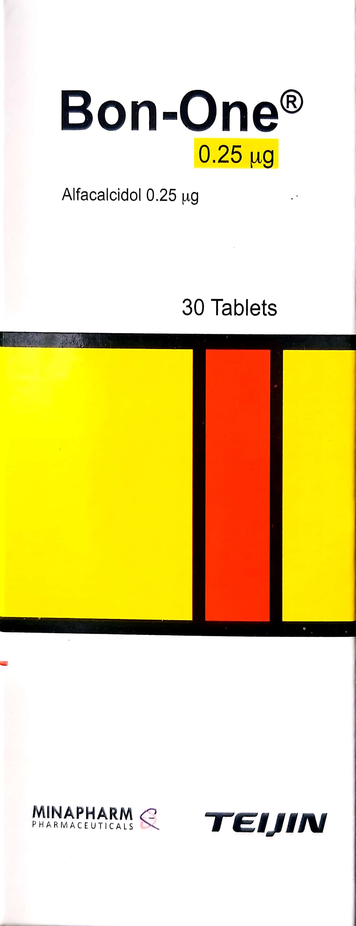
In a modern clinical setting, the management of a stable lumbar spinal compression fracture requires a multidisciplinary approach that integrates pharmacological, orthotic, and, when necessary, surgical interventions. For patients with underlying bone density issues, long-term management often includes Alendronate / ألندرونات 70 mg, Bon-one / بون-ون 0.25mcg, and Calcimed D3 Effervescent Tablets / أقراص كالسي ميد د3 الفوارة 600 mg Calcium / 400 IU Cholecalciferol to improve bone quality, while mechanical stabilization is achieved through the use of a TLSO Brace (Thoracolumbosacral Orthosis) / دعامة صدرية قطنية عجزية (TLSO) (الأطراف الصناعية والجبائر التقويمية). If conservative measures fail or pain persists, surgical stabilization may be indicated, utilizing procedures such as Kyphoplasty / رأب الحدباء (عملية كبرى في غرف العمليات) or Kyphoplasty/Vertebroplasty for Pathologic Oncologic Fracture / رأب الحدب/رأب الفقرات لكسر مرضي ورمي (عملية صغرى في العيادة). These procedures rely on specialized surgical instruments, including Laparoscopic Trocar (5mm, 10mm, 12mm) / مبزل منظار البطن (5 مم، 10 مم، 12 مم), [PMMA Bone Cement (Antibiotic Loaded) / إسمنت عظام PMMA (محمل بالمضادات الحيوية)](https://yemenhealthos.com/