Clinical Assessment & Protocol
Typical Presentation (HPI)
EN: Patient presents with acute onset of thoracic back pain following [minor trauma/mechanical stress]. Pain is localized to the [T-level] region, exacerbated by movement, flexion, and weight-bearing, and relieved by rest. Patient denies radicular symptoms, bowel/bladder dysfunction, or lower extremity weakness. No history of malignancy or constitutional symptoms. AR: يراجع المريض بسبب ألم حاد في الظهر في المنطقة الصدرية بعد [تعرض لرض خفيف/إجهاد ميكانيكي]. يتركز الألم في مستوى [T-level]، ويزداد مع الحركة والانحناء وحمل الأثقال، ويتحسن بالراحة. ينفي المريض وجود أعراض عصبية جذرية، أو خلل في الأمعاء/المثانة، أو ضعف في الأطراف السفلية. لا يوجد تاريخ مرضي للأورام أو أعراض جهازية.
General Examination
EN: Thoracic spine examination reveals localized tenderness to palpation over the [T-level] spinous process. No step-off deformity or paraspinal muscle spasm noted. Neurological examination is intact: motor strength 5/5 in all extremities, sensation intact to light touch, and deep tendon reflexes are 2+ and symmetric. Gait is antalgic but stable. AR: يكشف فحص العمود الفقري الصدري عن إيلام موضعي عند الجس فوق النتوء الشوكي للمستوى [T-level]. لا توجد تشوهات في المحاذاة أو تشنجات في العضلات المجاورة للفقرات. الفحص العصبي سليم: القوة الحركية 5/5 في جميع الأطراف، الإحساس سليم للمس الخفيف، والمنعكسات الوترية العميقة 2+ ومتناظرة. المشية متألمة ولكنها مستقرة.
Treatment Protocol
EN: Management plan includes conservative therapy: activity modification, avoidance of heavy lifting, and bracing (TLSO) as indicated for comfort. Analgesia initiated with [NSAIDs/Acetaminophen]. Referral to physical therapy for core strengthening once acute pain subsides. Follow-up imaging in [X] weeks to confirm stability. AR: تتضمن خطة العلاج تدابير تحفظية: تعديل النشاط، تجنب رفع الأثقال، واستخدام دعامة الظهر (TLSO) حسب الحاجة للراحة. تم البدء بمسكنات الألم [مضادات الالتهاب غير الستيرويدية/باراسيتامول]. تحويل المريض للعلاج الطبيعي لتقوية عضلات الجذع بعد زوال الألم الحاد. إجراء تصوير متابعة بعد [X] أسابيع للتأكد من استقرار الكسر.
Patient Education
EN: You have a stable compression fracture of the thoracic spine. This is a structural change in the bone that is not currently unstable. Focus on "log-rolling" when getting out of bed, avoid bending, lifting, or twisting. Wear your brace as directed. Seek immediate medical attention if you experience numbness, tingling, or loss of bowel/bladder control. AR: تعاني من كسر انضغاطي مستقر في الفقرات الصدرية. هذا تغير هيكلي في العظم وهو مستقر حالياً. ركز على تقنية "الدوران ككتلة واحدة" عند النهوض من السرير، وتجنب الانحناء أو رفع الأثقال أو الالتواء. ارتدِ الدعامة حسب التوجيهات. اطلب الرعاية الطبية الفورية إذا شعرت بتنميل، وخز، أو فقدان السيطرة على الأمعاء أو المثانة.
Systemic & Specialized Examinations
EN: Distinct radiculopathy (L4/L5/S1). Strict Cauda Equina precautions documented. AR: اعتلال عصبي جذري واضح. تم توثيق تحذيرات متلازمة ذيل الفرس بصرامة.
Orthopedic & Trauma Assessments
EN: Heavy lifting incident with spinal rotation, or insidious degenerative disc disease. AR: حادث رفع أوزان ثقيلة مع دوران للعمود الفقري، أو انزلاق غضروفي تدريجي.
EN: Antalgic gait. Exhibits a 'list' (sciatic scoliosis) away from the affected side. Difficulty with heel/toe walk. AR: مشية متألمة. يظهر ميلاً (جنف وركي) لتخفيف الضغط. صعوبة في المشي على الكعب/الأصابع.
EN: Loss of normal lumbar lordosis. Severe paraspinal muscle spasm. AR: فقدان التقوس القطني الطبيعي. تشنج عضلي شديد حول الفقرات.
EN: Straight Leg Raise (SLR): Strongly positive at 30-45°. Slump test positive. AR: اختبار رفع الساق المستقيمة (SLR): إيجابي بقوة عند 30-45 درجة.
EN: Weakness (4/5) in EHL (L5) or Plantarflexion (S1). AR: ضعف (4/5) في باسطة الإبهام (L5) أو الثني الأخمصي (S1).
EN: Hypoesthesia to pinprick over the foot dorsum (L5) or lateral border (S1). AR: نقص الإحساس للوخز على ظهر القدم (L5) أو الجانب الوحشي (S1).
EN: Achilles (S1) diminished 1+. Patellar (L4) 2+. AR: منعكس وتر أخيل ضعيف 1+. منعكس الرضفة طبيعي 2+.
EN: DP and PT pulses 2+ symmetric. AR: النبضات الطرفية طبيعية.
Clinical Comprehensive Guide: Stable Thoracic Spinal Compression Fracture
1. Introduction and Overview
A thoracic spinal compression fracture (TSCF) occurs when the vertebral body in the thoracic spine—the segment between the base of the neck and the abdomen—collapses due to axial loading or diminished bone density. A "stable" classification indicates that the posterior column of the vertebral unit remains intact, meaning there is no neurological compromise or risk of progressive deformity under normal physiological loads.
In clinical practice, these fractures represent a significant portion of orthopedic consultations, particularly in geriatric populations or trauma settings. While "stable" implies a lower immediate risk of spinal cord injury compared to unstable fractures (which involve the middle column, as defined by Denis’s Three-Column Theory), they still necessitate meticulous management to prevent chronic pain, kyphotic deformity, and secondary pulmonary complications.
2. Etiology and Pathophysiology
Etiology
The thoracic spine is uniquely susceptible to compression injuries due to its anatomical constraints and the presence of the rib cage. Etiologies are typically bifurcated into two primary categories:
- Osteoporotic/Insufficiency Fractures: The most common cause. Age-related bone mineral density (BMD) loss leads to micro-architectural deterioration, making the trabecular bone unable to withstand normal physiological stress.
- High-Energy Trauma: Occurs in younger, healthy populations due to motor vehicle accidents, falls from height, or contact sports involving hyperflexion of the torso.
- Pathological Fractures: Secondary to underlying malignancy (e.g., multiple myeloma, metastatic breast or prostate cancer) that weakens the vertebral structure.
Pathophysiology
The mechanical failure occurs primarily in the anterior column. When the compressive force exceeds the structural integrity of the vertebral body—specifically the cancellous bone—the anterior height is lost. Because the posterior ligaments (supraspinous, interspinous, and ligamentum flavum) and the posterior bony elements (pedicles, lamina, facets) remain intact, the vertebral column maintains its alignment, preventing translational instability.
| Feature | Stable TSCF | Unstable TSCF |
|---|---|---|
| Columns Involved | Anterior only | Middle and/or Posterior |
| Neurological Risk | Minimal | High (Cord compression) |
| Treatment | Conservative/Bracing | Surgical Stabilization |
| Deformity Risk | Low (if managed) | High (Kyphosis) |
3. Clinical Presentation and Staging
Standard Presentation
Patients typically present with localized, sharp thoracic pain exacerbated by movement, coughing, sneezing, or deep inspiration. In geriatric patients, the presentation may be subtle, manifesting as a "height loss" or a gradual increase in thoracic kyphosis (the "dowager’s hump").
- Physical Examination Findings:
- Point tenderness over the affected spinous process.
- Paravertebral muscle spasms.
- Limited spinal range of motion.
- Absence of focal neurological deficits (motor/sensory).
Clinical Staging (Genant Semi-Quantitative Method)
Clinicians often utilize the Genant scale to quantify the severity of the vertebral collapse:
- Grade 1 (Mild): 20–25% reduction in vertebral height.
- Grade 2 (Moderate): 25–40% reduction in vertebral height.
- Grade 3 (Severe): >40% reduction in vertebral height.
4. Differential Diagnosis
It is critical to rule out mimickers of stable TSCF, as the treatment pathways differ significantly:
- Vertebral Osteomyelitis: Presents with constitutional symptoms (fever, night sweats, elevated ESR/CRP).
- Spinal Metastasis: Often presents with unrelenting, nocturnal pain unresponsive to rest.
- Scheuermann’s Kyphosis: Developmental disorder, not a fracture, though often confused on imaging.
- Thoracic Disc Herniation: Typically involves radicular pain or dermatomal distribution, which is rare in pure compression fractures.
5. Diagnostic Testing Protocols
Imaging Modalities
- Plain Radiography (X-ray): First-line diagnostic tool. AP and lateral views are essential to assess height loss and alignment.
- Magnetic Resonance Imaging (MRI): The gold standard for determining the age of the fracture. A T2-weighted STIR (Short Tau Inversion Recovery) sequence will show high signal intensity (edema) in acute fractures.
- CT Scan: Used if there is suspicion of cortical breach or to evaluate the integrity of the posterior elements if X-rays are inconclusive.
- DEXA Scan: Mandatory for patients over 50 to assess for underlying osteoporosis.
6. Clinical Indications & Management Strategy
Conservative Management
The majority of stable TSCFs are managed non-operatively.
* Analgesia: Acetaminophen, NSAIDs, and in acute cases, a short course of calcitonin to reduce pain associated with bone turnover.
* Bracing: Thoracolumbar orthosis (TLSO) may be prescribed for comfort and to limit painful flexion, though evidence on long-term deformity prevention via bracing remains debated.
* Physical Therapy: Focus on gentle mobilization, core stabilization, and fall prevention training.
Indications for Intervention
While the fracture is "stable," surgical intervention (Kyphoplasty or Vertebroplasty) may be indicated if:
1. Pain is intractable despite 4-6 weeks of aggressive conservative management.
2. The patient suffers from pulmonary compromise due to severe kyphotic deformity.
3. The patient is experiencing significant decline in functional independence.
7. Risks, Side Effects, and Contraindications
Risks of Conservative Care
- Progressive Kyphosis: Even "stable" fractures can collapse further over time, leading to a fixed deformity.
- Pulmonary Restriction: Significant thoracic kyphosis can restrict lung expansion, reducing vital capacity.
- Secondary Fractures: The altered biomechanics of the spine increase the load on adjacent vertebrae, increasing the risk of subsequent fractures.
Contraindications for Surgical Intervention
- Active systemic infection or local soft tissue infection at the site.
- Coagulopathy that cannot be corrected.
- Fractures that are chronic/healed (where the bone is already sclerotic).
- Presence of tumor within the canal (requires different surgical approach).
8. Long-Term Prognosis
The prognosis for a stable TSCF is generally favorable. Most fractures heal within 8 to 12 weeks. However, the long-term prognosis is highly dependent on the management of the underlying bone density. Patients are at a high risk for a "fracture cascade," where one fracture leads to another. Long-term management must include anti-resorptive medications (bisphosphonates, denosumab) and lifestyle modifications to optimize bone health.
9. Comprehensive FAQ Section
1. Is a "stable" fracture safe to leave untreated?
While the spine is mechanically stable, "untreated" does not mean "ignored." Lack of pain management and physical therapy can lead to muscle atrophy and chronic pain. Medical supervision is required to ensure the fracture heals correctly.
2. How long does the pain last?
Acute pain typically subsides within 4–8 weeks. If pain persists beyond 3 months, it may indicate non-union or a new fracture.
3. Do I need surgery if I am in a lot of pain?
Not necessarily. Many patients find relief through a combination of bracing, physical therapy, and pain management. Surgery is generally reserved for those who fail to improve after 6 weeks.
4. Will I get shorter?
Some height loss is common with compression fractures. However, aggressive core strengthening and posture correction can minimize this effect.
5. Can I exercise with a stable TSCF?
Yes, but with modifications. Avoid high-impact activities, heavy lifting, and aggressive spinal flexion. Focus on low-impact activities like walking or stationary cycling.
6. What is the difference between Vertebroplasty and Kyphoplasty?
Vertebroplasty involves injecting cement into the collapsed bone. Kyphoplasty uses a balloon to restore height before injecting the cement.
7. Why is my doctor ordering an MRI for a fracture?
The MRI distinguishes between an acute (new) fracture and a chronic (old) one, which is vital for planning treatment.
8. Is a back brace mandatory?
Braces are used primarily for pain relief. Recent studies suggest that for many patients, the brace does not change the ultimate outcome of the fracture healing, but it provides significant comfort during the acute phase.
9. Can I drink alcohol or smoke after a fracture?
Smoking is strongly discouraged as it inhibits bone healing and reduces blood flow to the vertebrae. Alcohol in moderation is generally acceptable, but excessive intake increases fall risk.
10. What is the "Fracture Cascade"?
This refers to the clinical observation that patients who sustain one compression fracture are at a significantly higher risk of sustaining subsequent fractures in the thoracic or lumbar spine within the next 12 months.
10. Conclusion
A stable thoracic compression fracture is a manageable condition, but it serves as a critical sentinel event for overall skeletal health. The clinical priority is to stabilize the patient, manage pain, and aggressively treat the underlying metabolic bone disease. Through a multidisciplinary approach involving orthopedics, pain management, and physical therapy, patients can expect a return to their baseline functional status and a reduction in the risk of future spinal complications.
Related Clinical Integration
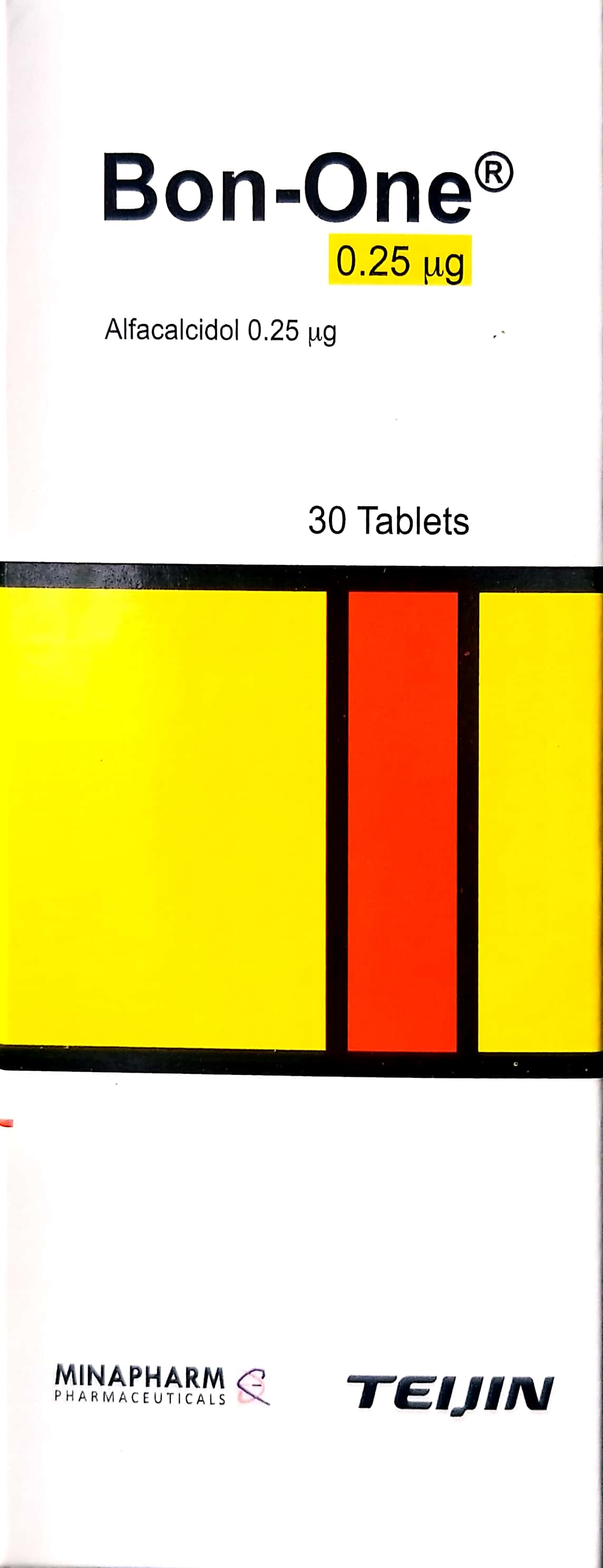
In the management of a stable thoracic spinal compression fracture, a multidisciplinary approach is essential to ensure structural stability, pain control, and long-term bone health. Clinical care often begins with conservative stabilization using a TLSO Brace (Thoracolumbosacral Orthosis) / دعامة صدرية قطنية عجزية (TLSO) (الأطراف الصناعية والجبائر التقويمية) to limit spinal motion, while pharmacological intervention focuses on bone density optimization through Alendronate / ألندرونات 70 mg, Bon-one / بون-ون 0.25mcg, and Calcimed D3 Effervescent Tablets / أقراص كالسي ميد د3 الفوارة 600 mg Calcium / 400 IU Cholecalciferol. Should the fracture fail to heal or present with underlying pathology, surgical intervention may be required, utilizing specialized equipment such as Bone Biopsy Needle Kit (e.g., Jamshidi, Trephine, Coaxial system) / مجموعة إبرة خزعة العظم (مثل: جامشيدي، تريفين، نظام محوري) for diagnostic sampling, Laparoscopic Trocar (5mm, 10mm, 12mm) / مبزل منظار البطن (5 مم، 10 مم، 12 مم) for access, and PMMA Bone Cement (Antibiotic Loaded) / إسمنت عظام PMMA (محمل بالمضادات الحيوية) for stabilization via [Kyphoplasty / رأب الحدباء (عملية كبرى في غرف العمليات)](https://yemenhealthos