Clinical Assessment & Protocol
Typical Presentation (HPI)
EN: Patient is an adolescent presenting with progressive varus deformity of the right knee. Symptoms include medial knee pain, intermittent limp, and cosmetic concerns regarding leg alignment. No history of acute trauma or infection. Symptoms exacerbated by physical activity. AR: المريض مراهق يعاني من تشوه تقوسي متزايد في الركبة اليمنى. تشمل الأعراض ألماً في الجانب الإنسي للركبة، عرجاً متقطعاً، ومخاوف تجميلية بشأن استقامة الساق. لا يوجد تاريخ لإصابة حادة أو عدوى. تزداد الأعراض سوءاً مع النشاط البدني.
General Examination
EN: Physical exam reveals right-sided genu varum with palpable medial tibial plateau prominence. Range of motion of the knee is full but with discomfort at terminal extension. Gait analysis demonstrates a varus thrust during the stance phase. Ligamentous stability is intact; no signs of joint effusion. AR: يكشف الفحص البدني عن وجود تقوس في الركبة اليمنى (genu varum) مع بروز ملموس في الهضبة الظنبوبية الإنسية. مدى حركة الركبة كامل ولكن مع وجود انزعاج عند التمديد النهائي. يظهر تحليل المشية وجود دفع تقوسي أثناء مرحلة الوقوف. استقرار الأربطة سليم؛ لا توجد علامات انصباب مفصلي.
Treatment Protocol
EN: Management plan includes radiographic assessment (long-leg standing films) to determine the mechanical axis and Langenskiöld stage. Treatment options discussed: weight management, physical therapy for gait optimization, and surgical intervention (guided growth or proximal tibial osteotomy) depending on skeletal maturity and severity of deformity. AR: تشمل خطة العلاج التقييم الشعاعي (صور الأشعة السينية للساق كاملة أثناء الوقوف) لتحديد المحور الميكانيكي ومرحلة لانجينسكولد (Langenskiöld stage). تمت مناقشة خيارات العلاج: إدارة الوزن، العلاج الطبيعي لتحسين المشية، والتدخل الجراحي (توجيه النمو أو قطع عظم الظنبوب القريب) بناءً على النضج الهيكلي وشدة التشوه.
Patient Education
EN: Blount's disease in adolescents is a growth disorder affecting the proximal tibia. It is important to maintain a healthy weight to reduce stress on the knee joint. Follow-up imaging is essential to monitor progression. Please report any increase in pain or change in gait immediately. AR: داء بلونت لدى المراهقين هو اضطراب في النمو يؤثر على الجزء القريب من عظم الظنبوب. من المهم الحفاظ على وزن صحي لتقليل الضغط على مفصل الركبة. التصوير المتابِع ضروري لمراقبة تطور الحالة. يرجى إبلاغنا فوراً في حال زيادة الألم أو حدوث أي تغير في المشية.
Systemic & Specialized Examinations
EN: Intact globally. AR: سليم.
Orthopedic & Trauma Assessments
EN: Developmental/Congenital etiology. No acute trauma. AR: سبب تطوري/خلقي. لا توجد صدمة حادة.
EN: Limping, toe-walking, or waddling gait observed (or pre-ambulatory infant). AR: يلاحظ عرج، مشي على الأصابع، أو مشية البطة (أو رضيع قبل مرحلة المشي).
EN: Asymmetric skin folds (gluteal/thigh). Apparent leg length discrepancy (Galeazzi sign positive). AR: طيات جلدية غير متماثلة (أرداف/فخذ). تباين واضح في طول الساقين (علامة غاليازي إيجابية).
EN: Barlow Maneuver: Provocative test reveals palpable clunk. Ortolani Maneuver: Gentle abduction reduces hip with clunk. AR: مناورة بارلو: تظهر طقطقة خلع. مناورة أورتولاني: ترد الورك بطقطقة.
EN: Moves all extremities equally. AR: يحرك جميع الأطراف بالتساوي.
EN: Withdraws to light stimulus. AR: يسحب الطرف استجابة للمس.
EN: 2+ symmetric. No clonus. AR: 2+ متماثلة.
EN: Strong and symmetric. AR: قوية ومتماثلة.
Comprehensive Clinical Guide: Adolescent Blount’s Disease (Tibia Vara), Right Knee
1. Comprehensive Introduction & Overview
Blount’s disease, clinically referred to as tibia vara, is a developmental disorder of the proximal tibial physis characterized by progressive multiplanar deformity of the knee. While infantile Blount’s disease is well-documented in toddlers, Adolescent Blount’s Disease (ABD)—also known as Late-Onset Tibia Vara—presents in children aged 10 years or older.
In the case of unilateral involvement, such as the right knee, the clinical presentation is often more subtle than the infantile form but significantly more complex regarding mechanical axial alignment. Adolescent Blount’s is strongly associated with obesity, rapid growth spurts, and repetitive mechanical stress on the medial proximal tibial physis. Unlike the infantile form, which often resolves with bracing, ABD is frequently progressive and typically requires surgical intervention to prevent long-term sequelae, including joint surface degeneration and chronic pain.
2. Deep-Dive: Etiology and Pathophysiology
The pathophysiology of Adolescent Blount’s disease is rooted in the "Heuter-Volkmann Law," which states that increased pressure across a physis inhibits longitudinal growth, while decreased pressure accelerates it.
The Mechanism of Deformity
- Mechanical Overload: The proximal medial tibial physis is subjected to excessive compressive forces, often exacerbated by a high Body Mass Index (BMI).
- Growth Arrest: Chronic compression leads to the premature closure or dysfunction of the medial proximal tibial physis.
- Asymmetric Growth: While the medial physis stops growing or slows down, the lateral physis continues to grow, resulting in the characteristic varus angulation (bowing) of the tibia.
- Internal Torsion: The deformity is rarely limited to the frontal plane. Patients often present with significant internal tibial torsion and procurvatum (anterior bowing) of the proximal tibia.
Risk Factors
- Obesity: The most significant correlate in adolescent cases.
- Early Puberty: Rapid skeletal growth velocity puts disproportionate stress on the physis.
- Mechanical Axis Deviation: Pre-existing ligamentous laxity or minor alignment issues that worsen with increased body mass.
3. Clinical Staging and Grading
Adolescent Blount’s is typically classified using the Langenskiöld Classification System, though it was originally designed for the infantile form; many clinicians now use the Scholefield and Barker classification specifically for late-onset disease.
Langenskiöld Stages (Modified for Adolescent Context)
| Stage | Radiographic Characteristics |
|---|---|
| I | Irregularity of the medial epiphyseal plate; widening of the physis. |
| II | Formation of a "beak" at the medial metaphysis. |
| III | Depression of the medial metaphysis; formation of a distinct bony bridge. |
| IV | Sharp, beak-like projection; significant medial depression. |
| V | Double-epiphyseal plate appearance; severe deformity. |
| VI | Complete osseous bridge across the physis; cessation of growth. |
4. Clinical Indications & Standard Presentation
Physical Examination Findings
- Varus Deformity: Visible bowing of the right knee when standing.
- Internal Tibial Torsion: The foot points inward while the patella may point forward or laterally.
- Joint Line Tenderness: Patients often report pain specifically along the medial joint line.
- Gait Abnormalities: An antalgic or "waddling" gait may be present, often with a compensatory lateral thrust of the knee during the stance phase of walking.
- Leg Length Discrepancy: If unilateral (right side), the right leg may appear shorter than the left due to the growth arrest.
Diagnostic Testing
- Standing Long-Leg Radiographs (Scanograms): The gold standard. Must include the hip, knee, and ankle to calculate the Mechanical Axis Deviation (MAD) and the Mechanical Lateral Distal Femoral Angle (mLDFA) and Medial Proximal Tibial Angle (MPTA).
- MRI: Essential to identify "physeal bar" formation (bony bridging) and to assess the integrity of the medial meniscus, which is often damaged in ABD.
- CT Scan: Utilized pre-operatively to map the precise location of the bony bridge if physeal bar resection is planned.
5. Differential Diagnosis
It is critical to distinguish Adolescent Blount’s from other causes of varus deformity to ensure appropriate treatment:
- Physiologic Genu Varum: Usually bilateral and self-correcting; does not present with the characteristic medial "beak" or metaphyseal depression.
- Rickets (Vitamin D Deficiency): Typically involves systemic signs (e.g., craniotabes, rachitic rosary) and diffuse metaphyseal cupping rather than localized medial involvement.
- Metaphyseal Chondrodysplasia: A systemic skeletal disorder that affects multiple growth plates, not just the proximal tibia.
- Post-Traumatic Growth Arrest: History of physeal fracture to the proximal tibia can mimic the radiographic appearance of Blount’s.
- Osteochondritis Dissecans: Can present with medial knee pain but does not cause the characteristic axial deformity of Blount’s.
6. Risks, Side Effects, and Contraindications
Risks of Untreated Disease
- Degenerative Arthritis: Chronic malalignment leads to premature wear of the medial compartment (medial compartment osteoarthritis).
- Ligamentous Instability: Chronic varus stress stretches the Lateral Collateral Ligament (LCL).
- Functional Limitation: Progressive difficulty with high-impact activities and chronic pain.
Surgical Risks
- Neurovascular Injury: Particularly the peroneal nerve during osteotomy/correction.
- Compartment Syndrome: A high risk in high-tibial osteotomies; requires vigilant post-operative monitoring.
- Non-union/Malunion: Failure of the bone to heal in the corrected position.
- Infection: Standard risks associated with internal or external fixation hardware.
Contraindications
- Active Infection: Osteomyelitis in the affected limb.
- Poor Nutritional Status: Compromises bone healing.
- Non-Compliance: Patients unable to follow weight-bearing restrictions are poor candidates for complex osteotomies.
7. Management and Prognosis
Non-Surgical Management
In the adolescent population, bracing is generally ineffective due to skeletal maturity and the high force vectors generated by increased body mass. Management is primarily focused on weight management and physical therapy to improve joint stability.
Surgical Management
- Guided Growth (Hemiepiphysiodesis): If the patient still has significant growth remaining, temporary tethering of the lateral physis (using an eight-plate) can encourage the medial side to catch up.
- High Tibial Osteotomy (HTO): The definitive treatment for severe deformity. The tibia is cut, realigned to restore the mechanical axis, and fixed with a plate or external fixator.
- Physeal Bar Resection: Rarely indicated in adolescents, but may be used if the bar is small and peripheral.
Long-Term Prognosis
With successful realignment, the prognosis is generally good. However, patients must be monitored for recurrence if growth remains. Long-term, these patients are at higher risk for early-onset osteoarthritis and may require joint arthroplasty in adulthood if the joint surface was significantly damaged prior to correction.
8. Massive FAQ Section
1. Is Adolescent Blount’s the same as Infantile Blount’s?
No. Infantile Blount’s occurs in toddlers and is often bilateral, while Adolescent Blount’s (late-onset) occurs in children >10, is often unilateral, and is strongly linked to obesity.
2. Can I use a brace to fix my right knee?
Generally, no. Bracing is rarely successful in adolescents because the bone is too rigid and the mechanical forces are too high.
3. Does obesity cause Blount’s disease?
Obesity is a major risk factor. The increased weight puts excessive pressure on the medial growth plate, causing it to stop growing prematurely.
4. Will I need surgery?
In most adolescent cases, surgery is required to correct the mechanical axis and prevent permanent joint damage.
5. What is an osteotomy?
An osteotomy is a surgical procedure where the bone is cut and realigned to a normal position and then held in place with hardware (plates/screws).
6. What is the "mechanical axis" and why does it matter?
The mechanical axis is a line drawn from the center of the hip to the center of the ankle. In a healthy knee, this line passes through the center of the knee. In Blount’s, it shifts medially, overloading the inner knee.
7. How long is the recovery from surgery?
Recovery typically involves 6–12 weeks of non-weight-bearing or restricted weight-bearing, followed by months of physical therapy.
8. Will the deformity come back after surgery?
If the patient still has significant growth remaining, there is a risk of recurrence, which is why surgeons often wait until the patient is closer to skeletal maturity.
9. Is physical therapy enough to correct the bowing?
Physical therapy can help with muscle strengthening and gait mechanics, but it cannot reverse the bony structural deformity caused by Blount’s.
10. What happens if I don't treat this?
Untreated Blount’s leads to progressive deformity, chronic pain, and early-onset osteoarthritis, often necessitating knee replacement at a young age.
9. Summary Table: Clinical Roadmap
| Phase | Action | Goal |
|---|---|---|
| Diagnosis | Standing X-rays + MRI | Confirm diagnosis and assess physeal bars. |
| Initial Management | Weight optimization | Reduce mechanical stress on the physis. |
| Surgical Planning | CT/Scanogram Analysis | Determine osteotomy site or guided growth strategy. |
| Post-Op | Physical Therapy | Regain range of motion and joint stability. |
| Follow-up | Annual Clinical Exam | Monitor for recurrence until skeletal maturity. |
Disclaimer: This guide is for educational purposes for healthcare professionals and students. Clinical decisions must always be individualized based on the patient’s specific radiological findings, skeletal age, and functional status. Consult with a fellowship-trained pediatric orthopedic surgeon for active cases.
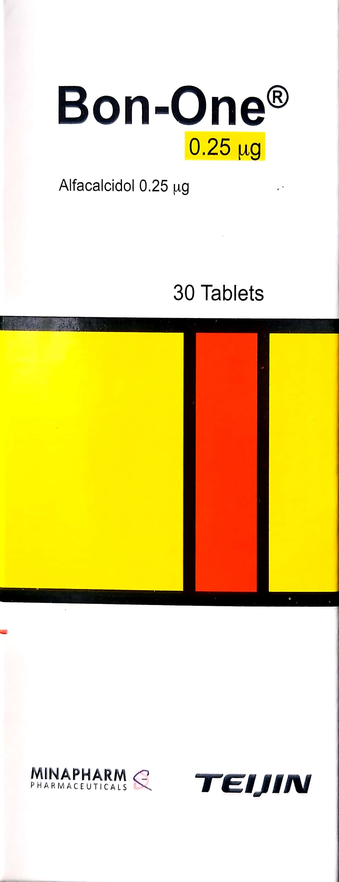
Related Clinical Integration
In the management of adolescent Blount’s Disease, a multidisciplinary approach is essential to address both the underlying metabolic factors and the structural deformity of the proximal tibia. Clinical care often begins with pharmacological support, utilizing Calcium Gluconate / غلوكونات الكالسيوم 10ml and Bon-one / بون-ون 0.25mcg to optimize bone health, while managing pain through non-steroidal anti-inflammatory drugs like Advil / أدفيل 200mg or Aleve / أليف 220mg. For patients requiring mechanical stabilization, orthotic interventions such as the Ankle Stirrup Brace / دعامة الكاحل على شكل ركاب (الأطراف الصناعية والجبائر التقويمية) or a Hip-Knee-Ankle-Foot Orthosis (HKAFO) / جبيرة الورك والركبة والكاحل والقدم (HKAFO) (الأطراف الصناعية والجبائر التقويمية) are utilized to offload the medial compartment. When surgical correction is indicated, orthopedic surgeons employ advanced instrumentation, including the Battery Powered Orthopedic Drill/Saw System / نظام مثقاب/منشار عظمي يعمل بالبطارية and the Guided Growth Plate (Eight-Plate) / صفيحة نمو موجهة (Eight-Plate), to perform corrective procedures such as a [Distal Femoral Osteotomy (DFO) / قطع العظم الفخذي القاصي (عملية كبرى في غرف العمليات)](https://yemenhealthos.com/ar/clinic/medical-procedures/distal-fem