Clinical Assessment & Protocol
Typical Presentation (HPI)
EN: Patient presents with chronic, progressive right knee pain, localized to the medial/lateral joint line, exacerbated by weight-bearing activities and prolonged standing. Reports morning stiffness lasting <30 minutes and occasional mechanical symptoms including crepitus and intermittent locking. AR: يعاني المريض من ألم مزمن ومتفاقم في الركبة اليمنى، يتركز في خط المفصل الإنسي/الوحشي، ويزداد سوءاً مع الأنشطة التي تتطلب تحميل الوزن والوقوف لفترات طويلة. يبلغ المريض عن تيبس صباحي يستمر لأقل من 30 دقيقة، مع أعراض ميكانيكية متقطعة تشمل الفرقعة (crepitus) والقفل المفصلي العرضي.
General Examination
EN: Patient is in no acute distress, ambulatory with a non-antalgic/antalgic gait. Systemic examination is unremarkable; patient is hemodynamically stable. AR: المريض في حالة عامة مستقرة ولا يبدو عليه ألم حاد، يمشي بمشية طبيعية/عرجاء. الفحص العام للجسم لا يظهر أي علامات غير طبيعية؛ المريض مستقر حيوياً.
Treatment Protocol
EN: Conservative management initiated: weight reduction, physical therapy for quadriceps strengthening, activity modification, and NSAIDs as needed. Intra-articular injection (corticosteroid/viscosupplementation) discussed. AR: تم البدء بالعلاج التحفظي: إنقاص الوزن، العلاج الطبيعي لتقوية العضلة الرباعية، تعديل الأنشطة، ومضادات الالتهاب غير الستيرويدية عند الحاجة. تمت مناقشة خيار الحقن داخل المفصل (كورتيكوستيرويد/مكملات لزوجة).
Patient Education
EN: Educated on the degenerative nature of osteoarthritis. Emphasized the importance of low-impact exercise, weight management, and adherence to physical therapy to maintain joint function and delay surgical intervention. AR: تم تثقيف المريض حول الطبيعة التنكسية لخشونة الركبة. تم التأكيد على أهمية ممارسة التمارين منخفضة التأثير، التحكم في الوزن، والالتزام بالعلاج الطبيعي للحفاظ على وظيفة المفصل وتأخير التدخل الجراحي.
Systemic & Specialized Examinations
EN: Unremarkable. Systemic examination is not the primary focus for this musculoskeletal pathology. AR: طبيعي. الفحص الجهازي ليس التركيز الأساسي لهذه الحالة العضلية الهيكلية.
EN: Unremarkable. Systemic examination is not the primary focus for this musculoskeletal pathology. AR: طبيعي. الفحص الجهازي ليس التركيز الأساسي لهذه الحالة العضلية الهيكلية.
EN: Unremarkable. Systemic examination is not the primary focus for this musculoskeletal pathology. AR: طبيعي. الفحص الجهازي ليس التركيز الأساسي لهذه الحالة العضلية الهيكلية.
EN: Unremarkable. Systemic examination is not the primary focus for this musculoskeletal pathology. AR: طبيعي. الفحص الجهازي ليس التركيز الأساسي لهذه الحالة العضلية الهيكلية.
EN: Unremarkable. Systemic examination is not the primary focus for this musculoskeletal pathology. AR: طبيعي. الفحص الجهازي ليس التركيز الأساسي لهذه الحالة العضلية الهيكلية.
EN: Unremarkable. Systemic examination is not the primary focus for this musculoskeletal pathology. AR: طبيعي. الفحص الجهازي ليس التركيز الأساسي لهذه الحالة العضلية الهيكلية.
EN: Unremarkable. Systemic examination is not the primary focus for this musculoskeletal pathology. AR: طبيعي. الفحص الجهازي ليس التركيز الأساسي لهذه الحالة العضلية الهيكلية.
EN: Unremarkable. Systemic examination is not the primary focus for this musculoskeletal pathology. AR: طبيعي. الفحص الجهازي ليس التركيز الأساسي لهذه الحالة العضلية الهيكلية.
EN: Unremarkable. Systemic examination is not the primary focus for this musculoskeletal pathology. AR: طبيعي. الفحص الجهازي ليس التركيز الأساسي لهذه الحالة العضلية الهيكلية.
Orthopedic & Trauma Assessments
EN: Insidious onset, consistent with primary degenerative osteoarthritis. No history of acute trauma or specific injury. AR: بداية تدريجية، تتوافق مع خشونة المفاصل التنكسية الأولية. لا يوجد تاريخ لصدمة حادة أو إصابة محددة.
EN: Gait is antalgic, favoring the right lower extremity with shortened stance phase on the affected side. AR: المشية عرجاء، مع تجنب تحميل الوزن على الطرف السفلي الأيمن وتقصير مرحلة الوقوف على الجانب المصاب.
EN: Right knee range of motion: Extension 0°, Flexion 110°. Terminal flexion limited by pain and osteophytic impingement. AR: مدى حركة الركبة اليمنى: البسط 0 درجة، الثني 110 درجات. الثني النهائي محدود بسبب الألم والنتوءات العظمية.
EN: Right knee inspection reveals mild joint line fullness, no gross deformity. Palpation confirms tenderness along the medial joint line. AR: فحص الركبة اليمنى يكشف عن امتلاء طفيف في خط المفصل، ولا توجد تشوهات ظاهرة. الجس يؤكد وجود إيلام على طول خط المفصل الإنسي.
EN: McMurray test negative for meniscal tear. Patellar grind test negative. AR: اختبار ماكموري سلبي لتمزق الغضروف الهلالي. اختبار طحن الرضفة سلبي.
EN: Motor strength 5/5 in quadriceps and hamstrings. No atrophy noted. AR: القوة العضلية 5/5 في العضلة الرباعية والعضلات المأبضية. لا يوجد ضمور عضلي.
EN: Sensation intact to light touch in all dermatomes of the right lower extremity. AR: الإحساس سليم للمس الخفيف في جميع القطاعات الجلدية للطرف السفلي الأيمن.
EN: Patellar and Achilles reflexes 2+ and symmetric. AR: منعكس الرضفة ومنعكس أخيل 2+ ومتماثلان.
EN: Dorsalis pedis and posterior tibial pulses 2+ and symmetric. AR: نبض الشريان ظهر القدم والشريان الظنبوبي الخلفي 2+ ومتماثلان.
Comprehensive Clinical Guide: Primary Osteoarthritis of the Right Knee
1. Introduction and Clinical Overview
Primary Osteoarthritis (OA) of the right knee, classified under ICD-10 code M17.11, represents a chronic, degenerative joint disease characterized by the progressive deterioration of articular cartilage, subchondral bone remodeling, and osteophyte formation. Unlike secondary osteoarthritis, which is triggered by specific trauma, congenital deformity, or systemic inflammatory conditions, primary OA is considered "idiopathic" or age-related.
The right knee is frequently a primary site of clinical complaint due to biomechanical loading patterns, repetitive micro-trauma, and the natural senescence of chondrocytes. As the most common form of arthritis globally, primary OA of the right knee results in significant morbidity, functional limitation, and decreased quality of life. This guide serves as an authoritative resource for understanding the multifaceted nature of this condition, from molecular pathophysiology to long-term clinical management.
2. Technical Specifications and Pathophysiology
The pathophysiology of primary OA is no longer viewed as a simple "wear and tear" phenomenon. It is now recognized as a complex, whole-joint failure involving the interplay of mechanical, metabolic, and inflammatory pathways.
The Molecular Cascade
- Chondrocyte Dysfunction: Chondrocytes, the sole cells in articular cartilage, undergo a phenotypic shift. They increase production of pro-inflammatory cytokines (IL-1β, TNF-α) and matrix-degrading enzymes (MMPs, ADAMTS).
- Matrix Degradation: The enzymatic breakdown of Type II collagen and aggrecan leads to the loss of the cartilage’s structural integrity, reducing its ability to withstand compressive forces.
- Subchondral Bone Remodeling: As cartilage thins, the subchondral bone experiences increased mechanical stress. This leads to bone sclerosis (thickening) and the formation of subchondral cysts.
- Synovial Inflammation: Chronic low-grade inflammation of the synovium (synovitis) exacerbates pain and further accelerates cartilage degradation through the release of inflammatory mediators into the synovial fluid.
Biomechanical Factors
The right knee, often the dominant leg in many patients, is subject to high peak forces during the stance phase of gait. Malalignment—specifically varus (bow-legged) or valgus (knock-kneed) deformity—shifts the center of gravity, causing uneven distribution of force across the medial or lateral compartments, respectively.
3. Clinical Staging and Grading
The most widely utilized clinical tool for assessing the severity of primary OA of the right knee is the Kellgren-Lawrence (K-L) Grading Scale.
| Grade | Classification | Radiographic Features |
|---|---|---|
| 0 | None | No radiographic features of OA. |
| 1 | Doubtful | Questionable osteophyte formation. |
| 2 | Mild | Definite osteophytes, unimpaired joint space. |
| 3 | Moderate | Moderate joint space narrowing. |
| 4 | Severe | Severe joint space narrowing, subchondral sclerosis. |
4. Clinical Presentation and Diagnostic Criteria
Standard Presentation
- Pain: Typically localized to the medial or anterior aspect of the right knee. Pain is usually exacerbated by activity (weight-bearing) and relieved by rest.
- Morning Stiffness: Generally lasts less than 30 minutes.
- Mechanical Symptoms: Patients may report catching, locking, or "giving way," often indicative of loose bodies or meniscal fraying secondary to OA.
- Crepitus: Audible or palpable grating during joint movement.
- Range of Motion (ROM) Deficits: Progressive loss of flexion and extension, often leading to a flexion contracture.
Diagnostic Workup
To confirm a diagnosis of primary OA of the right knee, the American College of Rheumatology (ACR) clinical criteria are often applied:
1. Knee pain.
2. Osteophytes on radiographs.
3. Synovial fluid analysis (if effusion is present) showing non-inflammatory characteristics.
4. Patient age > 50 years.
5. Morning stiffness < 30 minutes.
6. Crepitus on active motion.
5. Differential Diagnosis
Distinguishing primary OA from other pathologies is critical for effective treatment.
- Rheumatoid Arthritis (RA): Typically bilateral, symmetric, and characterized by prolonged morning stiffness (>60 mins) and systemic markers (RF, Anti-CCP).
- Meniscal Tear: Presents with acute pain and locking; clinical examination (McMurray’s test) is usually positive.
- Patellofemoral Pain Syndrome (PFPS): Pain primarily at the anterior knee, often in younger patients; radiographic findings of OA are absent.
- Gout/Pseudogout: Acute, intense inflammatory episodes; synovial fluid analysis confirms crystals (uric acid or calcium pyrophosphate).
- Septic Arthritis: Medical emergency; presents with fever, erythema, and severe pain; requires urgent aspiration.
6. Risks, Side Effects, and Contraindications
Risks of Advanced OA
- Functional Disability: Significant gait deviations leading to secondary hip or lumbar spine pain.
- Muscle Atrophy: Quadriceps inhibition (arthrogenic muscle inhibition) leads to instability.
Contraindications for Conservative Treatment
- NSAID Use: Contraindicated in patients with history of GI bleeds, severe renal impairment, or uncontrolled hypertension.
- Corticosteroid Injections: Contraindicated in the presence of suspected joint infection or skin breakdown over the injection site.
Surgical Risks (Arthroplasty)
- Periprosthetic infection.
- Deep Vein Thrombosis (DVT) and Pulmonary Embolism (PE).
- Implant loosening or mechanical failure.
7. Management Strategy
Treatment is hierarchical, moving from non-pharmacological to surgical interventions.
- First-Line: Patient education, weight management, and physical therapy (targeting quadriceps strengthening).
- Second-Line: Topical NSAIDs, oral acetaminophen/NSAIDs, and bracing (unloader braces for varus deformity).
- Third-Line: Intra-articular injections (corticosteroids, Hyaluronic Acid, or PRP).
- Final-Line: Total Knee Arthroplasty (TKA) or Unicompartmental Knee Arthroplasty (UKA).
8. Frequently Asked Questions (FAQ)
1. Is there a way to reverse primary osteoarthritis?
Currently, there is no cure for OA. Treatment focuses on symptom management, functional improvement, and slowing the rate of degradation.
2. Does weight loss actually help the right knee?
Yes. Every pound of body weight lost reduces approximately 4 pounds of force across the knee joint during each step, significantly reducing pain and inflammation.
3. Should I avoid exercise if I have knee pain?
No. Activity avoidance leads to muscle atrophy and stiffness. Low-impact exercises like swimming, cycling, and walking are highly recommended.
4. What is the difference between an X-ray and an MRI for OA?
X-rays are the gold standard for grading OA. MRIs are generally not required unless there is suspicion of soft tissue injury (e.g., ligament or meniscus tear).
5. Are dietary supplements like Glucosamine effective?
Clinical evidence is mixed. While some patients report relief, large-scale studies have not consistently shown superiority over placebo for structural cartilage regeneration.
6. Will I eventually need a knee replacement?
Not necessarily. Many patients manage primary OA successfully for years through physical therapy and lifestyle modifications. Surgery is reserved for those who remain symptomatic despite exhaustive conservative care.
7. How long does a knee replacement last?
Modern implants typically last 15–20 years, depending on the patient's activity level and weight.
8. Why is my right knee clicking or popping?
This is usually caused by crepitus (bone-on-bone friction) or the movement of irregular cartilage surfaces. It is generally not concerning unless accompanied by acute pain or locking.
9. Can weather changes affect my knee pain?
Yes. Changes in barometric pressure can cause expansion or contraction of the joint capsule and surrounding tissues, leading to increased pain in sensitive, arthritic joints.
10. When should I seek an immediate orthopedic evaluation?
If you experience sudden swelling, inability to bear weight, visible deformity, or symptoms of infection (fever, redness, warmth), you should seek immediate care.
9. Long-term Prognosis
The prognosis for primary OA of the right knee is variable. While the condition is progressive, the rate of progression is highly individualized. Patients who engage in structured exercise programs, maintain a healthy BMI, and manage co-morbidities generally experience better outcomes and delay the need for surgical intervention.
Early diagnosis and the adoption of a multidisciplinary care approach—involving orthopedists, physical therapists, and pain management specialists—are the most effective strategies for maintaining long-term joint health and mobility. Primary OA of the right knee should not be viewed as a signal of inevitable disability, but as a condition that requires proactive, evidence-based management to preserve function throughout the aging process.
Disclaimer: This guide is intended for informational purposes for healthcare professionals and patients. It does not replace professional medical advice, diagnosis, or treatment. Always consult with a qualified orthopedic specialist for individual care plans.
Related Clinical Integration
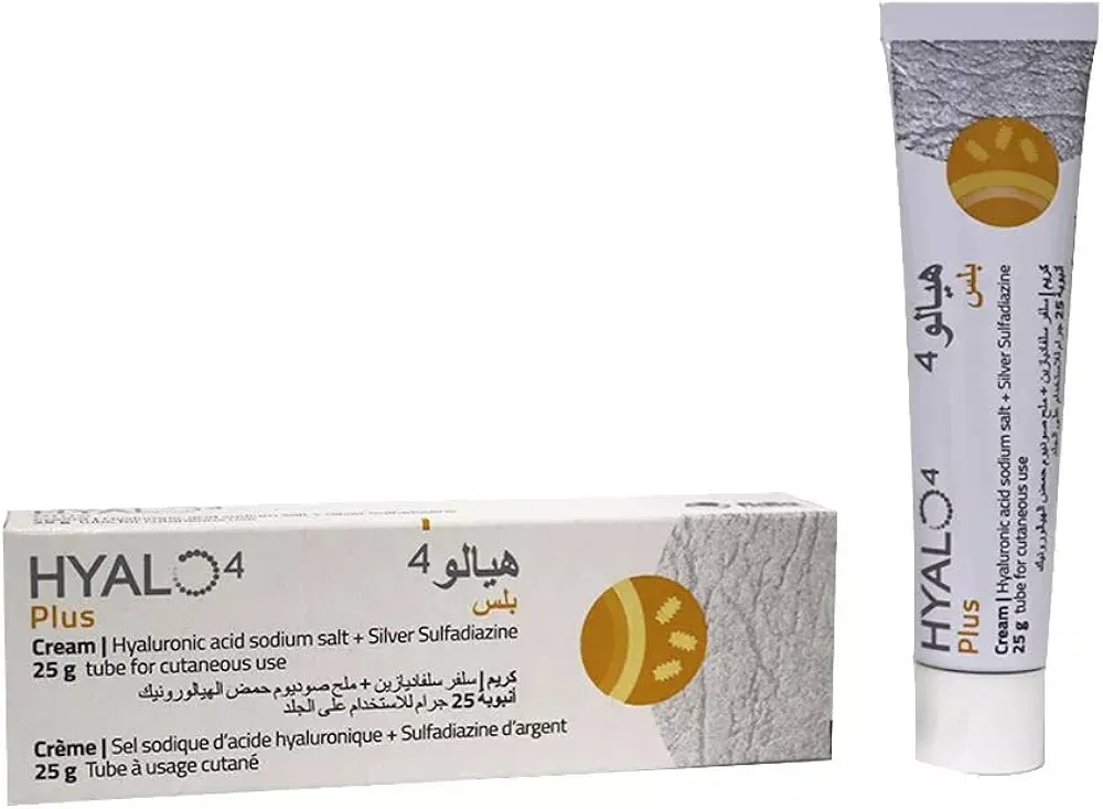
In the management of Primary Osteoarthritis of the Right Knee, a multidisciplinary clinical approach is essential to optimize patient outcomes across the continuum of care. Initial conservative management often involves pharmacological interventions such as Celcox / سيلكوكس 100mg, Meloxicam / ميلوكسيكام 25mg, and topical agents like Hyalo4 plus cream / هيالو 4 بلس كريم 0.2% / 1% to mitigate inflammation and pain. When non-operative measures fail to provide relief, surgical intervention may be indicated, necessitating advanced procedures such as Total Knee Arthroplasty (TKA) / استبدال مفصل الركبة الكلي (TKA) (عملية كبرى في غرف العمليات), which relies on precision tools like the Oscillating Bone Saw Blade (Wide, Narrow, Deep Cut) / شفرة منشار عظمي متذبذب (عريض، ضيق، قطع عميق). To ensure evidence-based practice, clinicians should consult foundational resources including Knee Arthritis: Epidemiology, Pathophysiology, Diagnosis & Surgical Anatomy, Primary Tricompartmental Knee Replacement: Advanced Surgical Technique, and Primary Total Knee Arthroplasty: Functional Outcomes, Radiographic Evaluation, and Long-Term Survivorship. Furthermore, complex presentations may require review of Advanced Bilateral Knee Osteoarthritis: A Comprehensive Clinical Case Study or specialized procedures