Clinical Assessment & Protocol
Typical Presentation (HPI)
EN: Chronic medial knee pain exacerbated by weight-bearing and stairs. AR: ألم مزمن في الركبة الإنسية يزداد مع تحميل الوزن وصعود الدرج.
General Examination
EN: Unremarkable or not routinely indicated. AR: طبيعي أو غير مطلوب روتينياً.
Treatment Protocol
EN: Quadriceps strengthening, bracing, and weight management. AR: تقوية العضلة الرباعية، استخدام دعامات، وإدارة الوزن.
Patient Education
EN: Activity modification and impact reduction strategies. AR: تعديل الأنشطة واستراتيجيات تقليل الصدمات.
Systemic & Specialized Examinations
EN: S1, S2 present. No murmurs. AR: صوتا القلب الأول والثاني طبيعيان. لا توجد نفخات.
EN: Lungs clear to auscultation. AR: الرئتان صافيتان عند التسمع.
EN: Abdomen soft, non-tender. AR: البطن لين ولا يوجد ألم.
EN: Alert, oriented x3. No focal deficits. AR: المريض واعي ومدرك. لا يوجد عجز عصبي بؤري.
EN: Unremarkable or not routinely indicated. AR: طبيعي أو غير مطلوب روتينياً.
EN: Unremarkable or not routinely indicated. AR: طبيعي أو غير مطلوب روتينياً.
EN: Unremarkable or not routinely indicated. AR: طبيعي أو غير مطلوب روتينياً.
EN: Unremarkable or not routinely indicated. AR: طبيعي أو غير مطلوب روتينياً.
EN: Unremarkable or not routinely indicated. AR: طبيعي أو غير مطلوب روتينياً.
Orthopedic & Trauma Assessments
EN: Joint line tenderness, crepitus on flexion, and varus malalignment. AR: إيلام عند خط المفصل، فرقعة عند الثني، وتشوه في المحاذاة (تقوس).
EN: Unremarkable or not routinely indicated. AR: طبيعي أو غير مطلوب روتينياً.
Comprehensive Clinical Guide: Medial Compartment Knee Osteoarthritis (MCOA)
1. Comprehensive Introduction & Overview
Medial Compartment Osteoarthritis (MCOA) represents the most prevalent form of knee osteoarthritis (OA), a progressive, degenerative condition characterized by the mechanical wear and degradation of articular cartilage within the medial tibiofemoral joint. Unlike global knee osteoarthritis, which involves all three compartments (medial, lateral, and patellofemoral), MCOA is localized, often driven by biomechanical malalignment and repetitive axial loading.
The medial compartment naturally bears a significantly higher percentage of body weight compared to the lateral compartment during the gait cycle. Consequently, when structural integrity is compromised—whether through trauma, obesity, or constitutional varus alignment—the medial compartment becomes the primary site of cartilage erosion, subchondral bone remodeling, and secondary inflammatory response. This guide serves as an authoritative resource for clinicians, medical students, and specialized healthcare providers to understand the nuances of diagnosing and managing MCOA.
2. Technical Specifications & Pathophysiology
The pathology of MCOA is not merely "wear and tear"; it is a complex, active biological process involving the entire joint organ, including the subchondral bone, synovium, and meniscus.
The Mechanical Axis
The normal mechanical axis of the lower limb passes near the center of the knee. In MCOA, the axis often shifts medially. This shift increases the adduction moment at the knee, creating a positive feedback loop:
1. Initial Varus Alignment: Increases stress on the medial condyle.
2. Cartilage Degradation: Proteoglycan loss and collagen network disruption.
3. Subchondral Bone Sclerosis: Increased bone density leads to decreased shock absorption, accelerating cartilage wear.
4. Osteophyte Formation: The body attempts to increase joint surface area to distribute load, resulting in bony outgrowths.
Pathophysiological Stages
| Phase | Biological Characteristic | Clinical Correlation |
|---|---|---|
| Early | Synovial inflammation, cartilage softening (chondromalacia). | Intermittent pain after activity. |
| Mid | Fibrillation of cartilage, micro-fractures in subchondral bone. | Persistent pain, stiffness, mild swelling. |
| Late | Full-thickness cartilage loss, bone-on-bone contact. | Severe pain, deformity, restricted range of motion. |
3. Clinical Staging: The Kellgren-Lawrence (KL) System
The KL system remains the gold standard for classifying the radiographic severity of MCOA.
- Grade 0 (None): No radiographic features of OA.
- Grade 1 (Doubtful): Doubtful joint space narrowing (JSN) and possible osteophytic lipping.
- Grade 2 (Mild): Definite osteophytes and possible JSN.
- Grade 3 (Moderate): Multiple osteophytes, definite JSN, sclerosis, and possible deformity of bone ends.
- Grade 4 (Severe): Large osteophytes, marked JSN, severe sclerosis, and definite deformity of bone ends (bone-on-bone).
4. Clinical Indications & Standard Presentation
Patients with MCOA typically present with a distinct clinical profile. Early identification is crucial for non-surgical intervention.
Key Symptoms:
- Medial Joint Line Tenderness: Localized pain upon palpation along the medial tibiofemoral joint line.
- Activity-Related Pain: Pain that worsens with weight-bearing activities (walking, stairs) and improves with rest.
- Stiffness: Morning stiffness lasting <30 minutes.
- Crepitus: Audible or palpable grinding sensation during flexion/extension.
- Varus Deformity: Visible "bow-legged" appearance in advanced stages.
Physical Examination Maneuvers:
- Varus Stress Test: Assessing stability of the medial collateral ligament (MCL).
- McMurray Test: Specifically looking for secondary medial meniscal pathology.
- Gait Analysis: Observation of a "medial thrust" or lateral trunk lean to compensate for varus loading.
5. Differential Diagnosis
Clinicians must distinguish MCOA from other conditions that mimic medial-sided knee pain:
* Medial Meniscal Tear: Often acute onset, mechanical locking or catching.
* Pes Anserine Bursitis: Point tenderness distal to the joint line (medial tibial plateau).
* Medial Collateral Ligament (MCL) Strain: History of trauma, laxity on valgus stress.
* Osteonecrosis of the Medial Femoral Condyle: Sudden onset, severe pain, often seen in older, sedentary patients (Knee SPON).
* Referred Pain: Hip pathology (e.g., hip OA) frequently presents as knee pain.
6. Diagnostic Testing Protocols
A systematic approach ensures diagnostic accuracy:
- Radiography (Weight-Bearing):
- AP View: Essential for assessing JSN.
- Rosenberg View (45-degree flexion PA): Superior for detecting early joint space narrowing that standard AP views might miss.
- Lateral View: To rule out patellofemoral involvement.
- MRI: Not required for routine OA diagnosis but indicated if:
- Mechanical symptoms suggest a meniscal flap tear.
- Bone marrow edema is suspected (a major pain generator).
- Surgical planning is underway.
- Laboratory Blood Work: Generally normal. Used only to rule out inflammatory arthropathies (e.g., Rheumatoid Arthritis, Gout) if the clinical presentation is atypical (e.g., bilateral involvement, systemic symptoms).
7. Risks, Side Effects, and Contraindications
Management strategies carry inherent risks that must be discussed with patients:
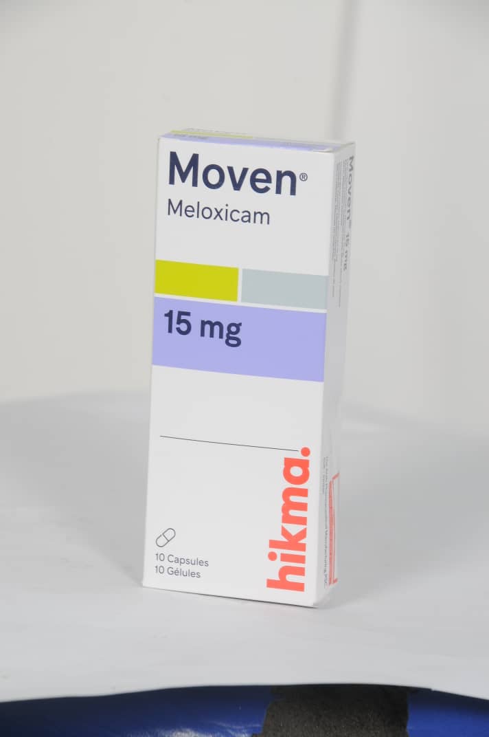
- NSAIDs: Risk of gastrointestinal bleeding, renal impairment, and hypertension. Contraindicated in patients with history of peptic ulcer disease or severe CHF.
- Corticosteroid Injections: Potential for cartilage toxicity if overused; strictly limited to 3-4 per year.
- Hyaluronic Acid (Viscosupplementation): Local reaction, post-injection flare.
- Surgical Intervention (High Tibial Osteotomy/TKA): Infection, DVT, pulmonary embolism, nerve injury, and persistent pain.
8. Long-Term Prognosis
MCOA is a chronic, degenerative condition. While it cannot be "cured," it can be managed effectively.
* Prognostic Factors for Progression: High BMI, baseline severity (KL grade), and poor quadriceps strength.
* Long-term Outcomes: With conservative care (physiotherapy, weight loss), many patients maintain function for years. Surgical intervention (Total Knee Arthroplasty) remains the definitive endpoint for end-stage (KL Grade 4) disease, boasting a 90%+ success rate in pain reduction and functional restoration.
9. FAQ Section (10 Frequently Asked Questions)
Q1: Can MCOA be reversed with supplements like Glucosamine?
A: Clinical evidence is mixed. While some patients report symptomatic relief, there is currently no high-level evidence that these supplements can regenerate lost cartilage or reverse the structural changes of MCOA.
Q2: Why does my knee hurt more when it rains?
A: While scientific consensus is evolving, barometric pressure drops can cause expansion of soft tissues and joint fluid, increasing pressure on sensitized nerve endings within an arthritic joint.
Q3: Is exercise dangerous for someone with MCOA?
A: No. Inactivity leads to muscle atrophy, which reduces joint stability and increases pain. Low-impact, high-repetition exercises (cycling, swimming) are highly recommended.
Q4: When is surgery absolutely necessary?
A: Surgery is considered when conservative measures (PT, weight loss, bracing, injections) fail to provide an acceptable quality of life, or when structural deformity significantly impairs mobility.
Q5: Will a knee brace help my medial compartment pain?
A: Yes. An "offloader" brace is designed to shift the weight-bearing load from the damaged medial compartment to the healthier lateral compartment.
Q6: Is weight loss really that important?
A: Crucially so. Every pound of body weight equates to approximately 4 pounds of force on the knee. A 10% reduction in body weight can lead to a 20-30% reduction in knee pain.
Q7: Can I continue running with MCOA?
A: High-impact running is generally discouraged for severe OA. Low-impact alternatives are preferred to preserve the remaining articular surface.
Q8: Are injections (PRP/Stem Cell) a standard treatment?
A: These are considered "orthobiologics." While emerging, they are not yet universally covered by insurance and are considered experimental by many medical boards.
Q9: Does MCOA always lead to total knee replacement?
A: No. Many patients manage their symptoms successfully for decades through lifestyle modifications and conservative therapy without ever requiring surgery.
Q10: What is the "medial thrust" gait?
A: This is a visible shift of the knee toward the midline during the stance phase of walking. It is a hallmark sign of medial compartment overload and indicates an abnormal biomechanical load on the joint.
10. Clinical Summary Table: Management Hierarchy
| Modality | Recommendation Strength | Goal |
|---|---|---|
| Weight Loss | Strong | Reduce mechanical load. |
| Quadriceps Strengthening | Strong | Dynamic stabilization of the joint. |
| Offloader Bracing | Moderate | Compartment-specific load redistribution. |
| NSAIDs (Topical/Oral) | Strong | Anti-inflammatory/Analgesic. |
| Corticosteroid Injection | Moderate | Short-term flare management. |
| Total Knee Arthroplasty | Strong (for End-Stage) | Restoration of function/pain relief. |
Disclaimer: This document is for educational purposes for healthcare professionals and does not replace professional clinical judgment or institutional protocols. Always perform an individualized assessment for each patient.